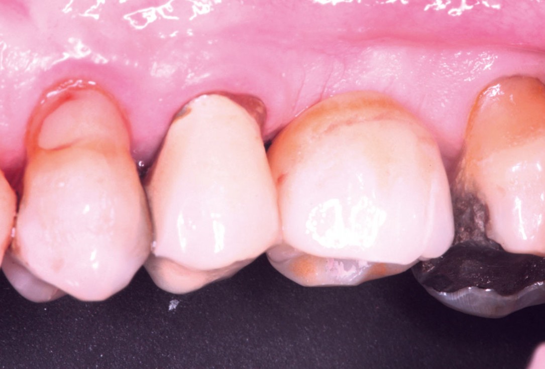
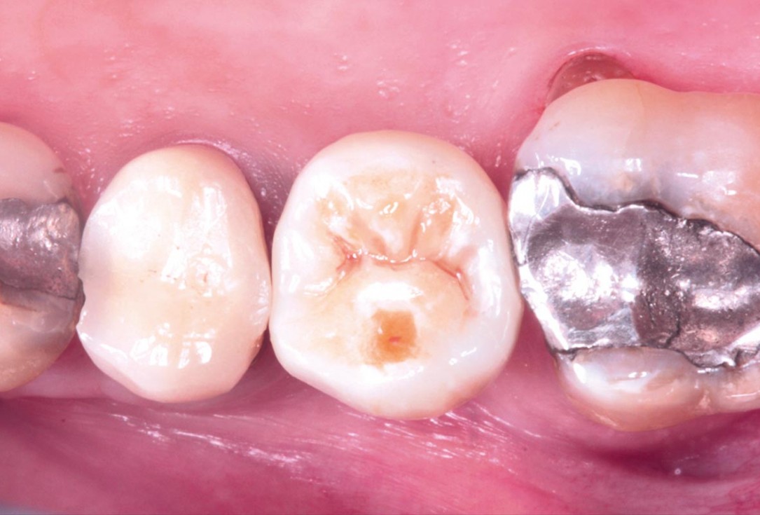
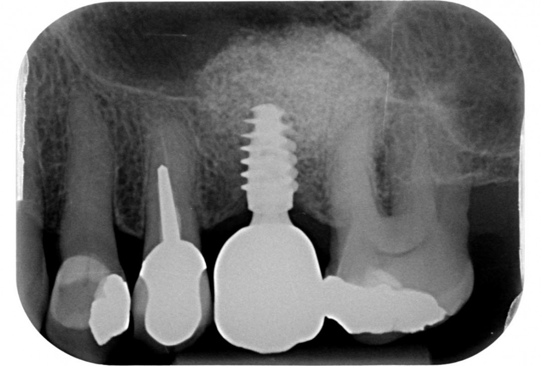
Maxillary sinus cyst removal using the Crocodile Technique and subsequent lateral sinus lift - Dres. C. Scognamiglio and A. Perucchi
-
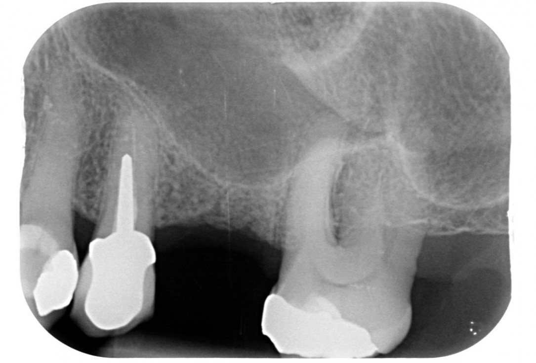
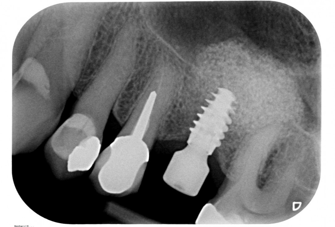
01/35 - Initial x-ray, tooth 25 compromised and to be extractedMaxillary sinus cyst removal using the Crocodile Technique and subsequent lateral sinus lift - Dres. C. Scognamiglio and A. Perucchi
-
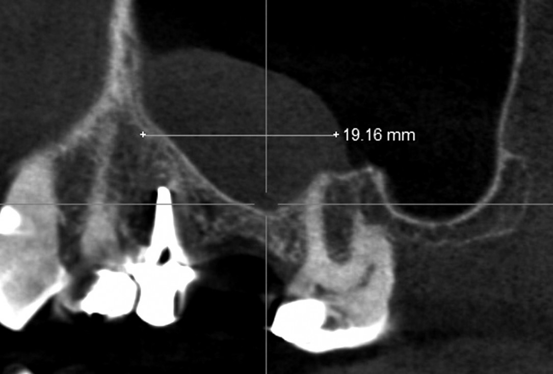
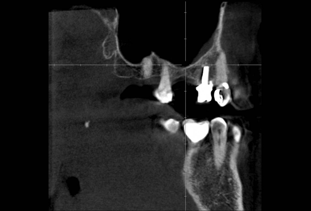
02/35 - CBCT shows maxillary sinus cystMaxillary sinus cyst removal using the Crocodile Technique and subsequent lateral sinus lift - Dres. C. Scognamiglio and A. Perucchi
-
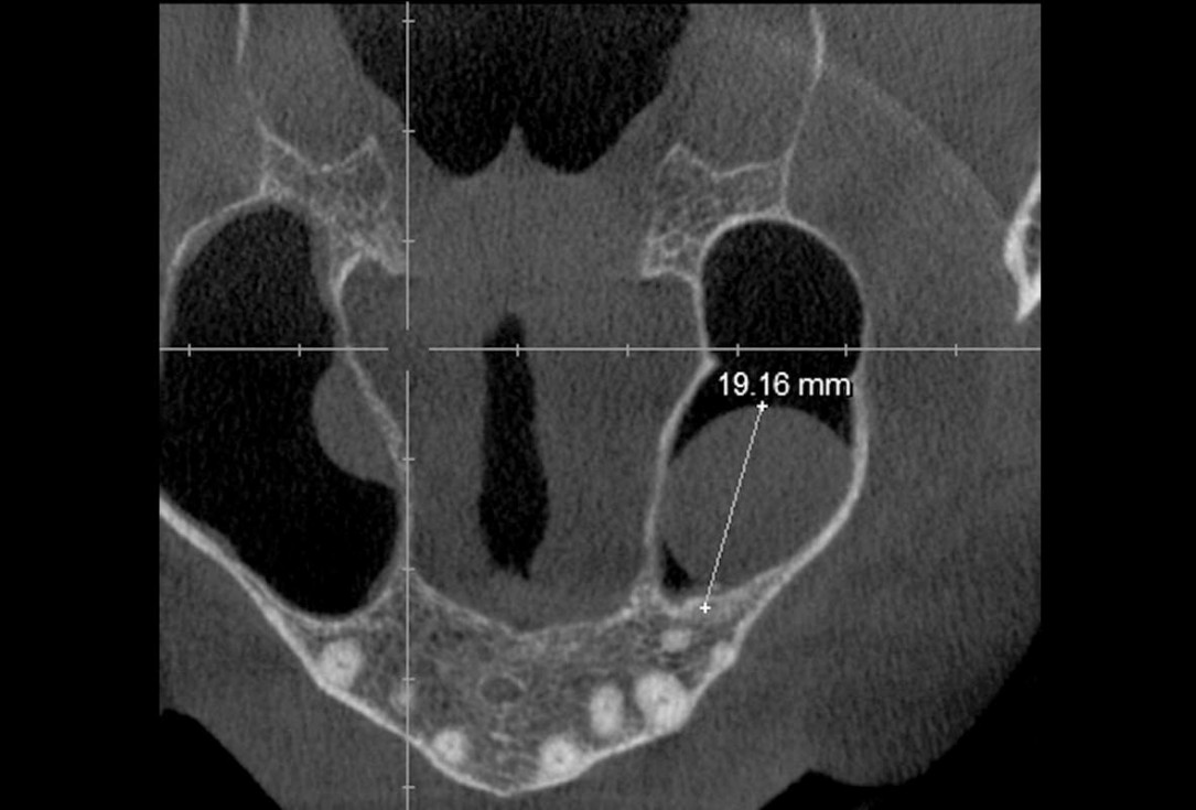
03/35 - CBCT shows maxillary sinus cystMaxillary sinus cyst removal using the Crocodile Technique and subsequent lateral sinus lift - Dres. C. Scognamiglio and A. Perucchi
-
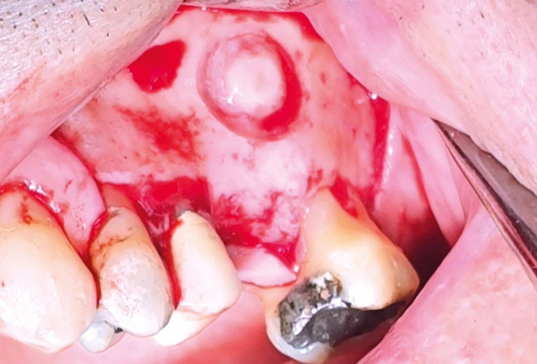
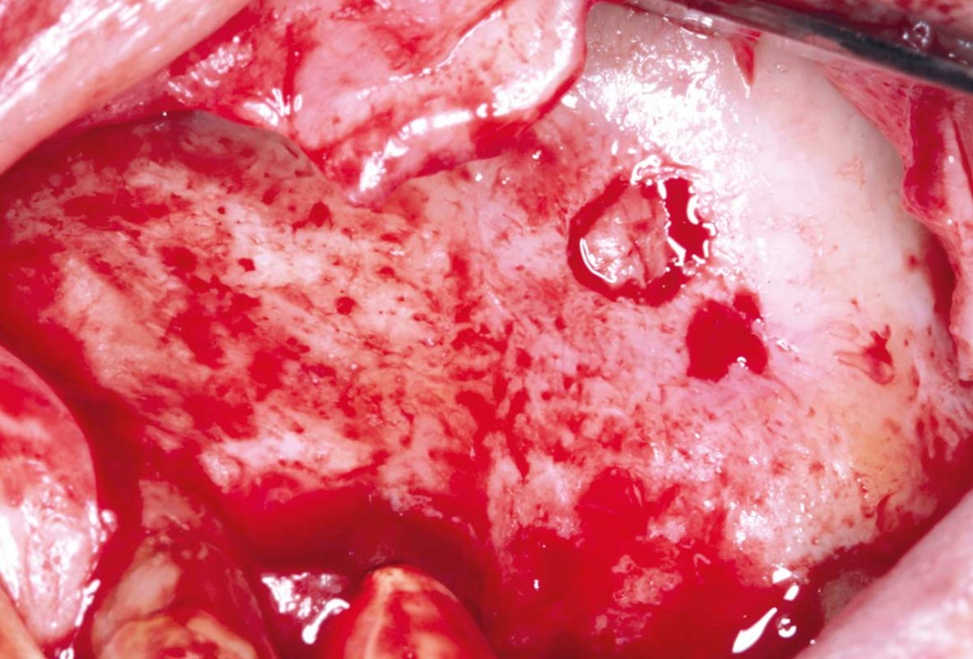
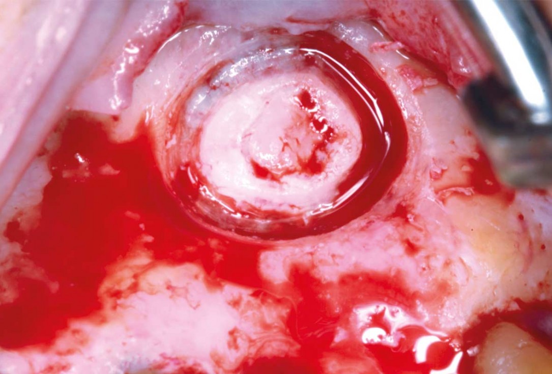
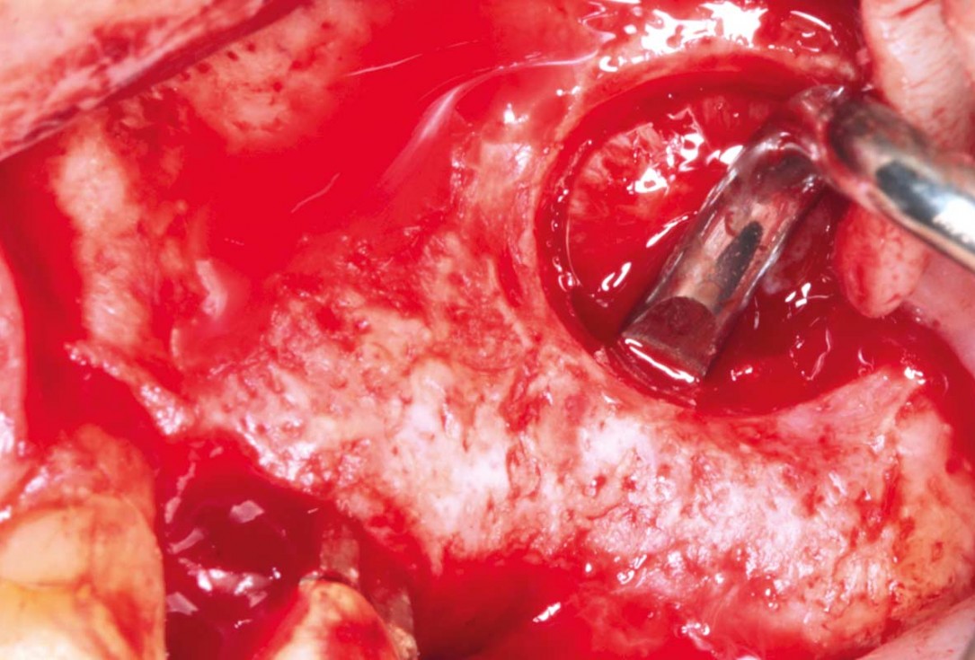
04/35 - Preparation of the lateral sinus window using piezosurgeryMaxillary sinus cyst removal using the Crocodile Technique and subsequent lateral sinus lift - Dres. C. Scognamiglio and A. Perucchi
-
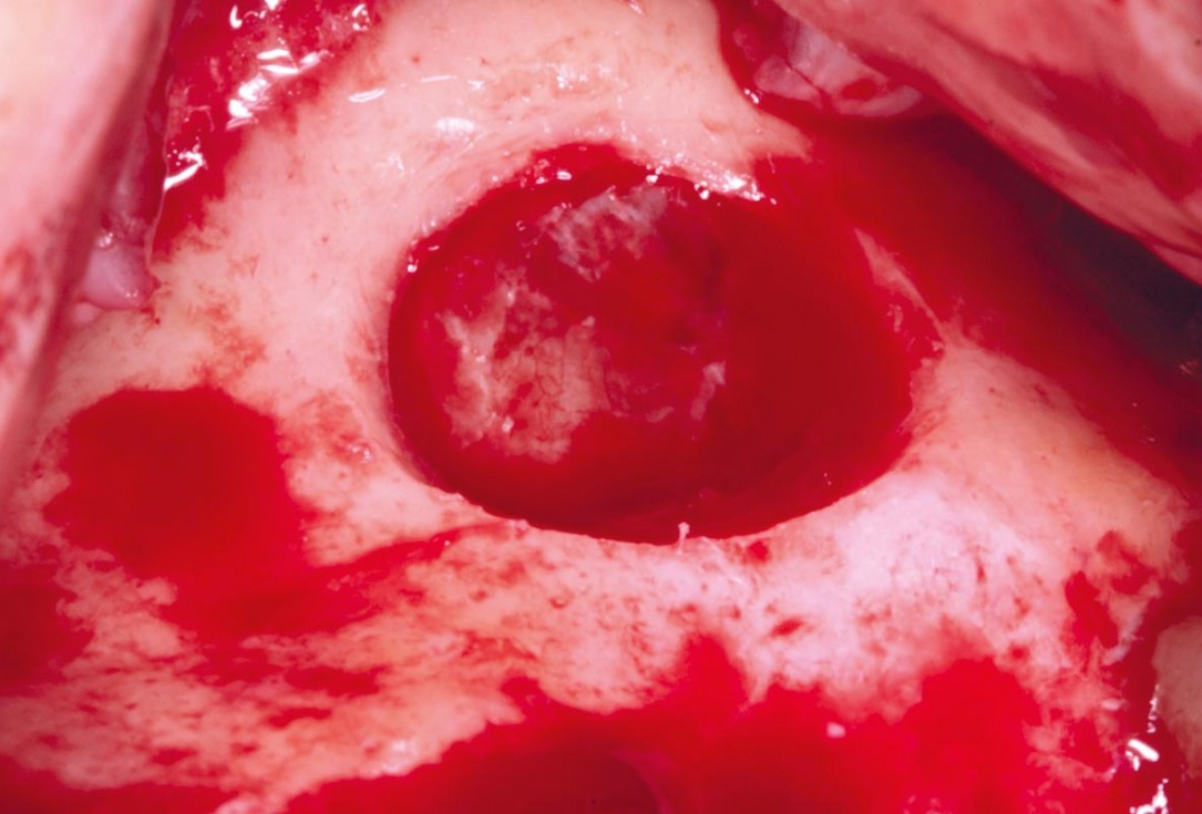
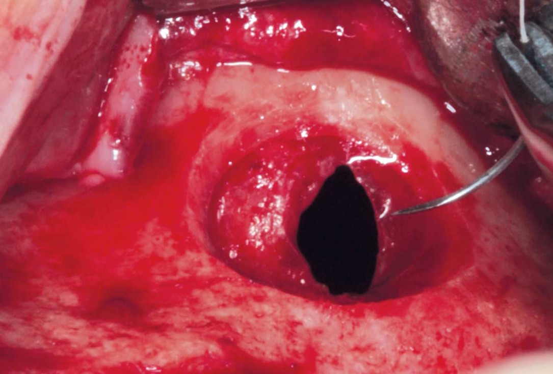
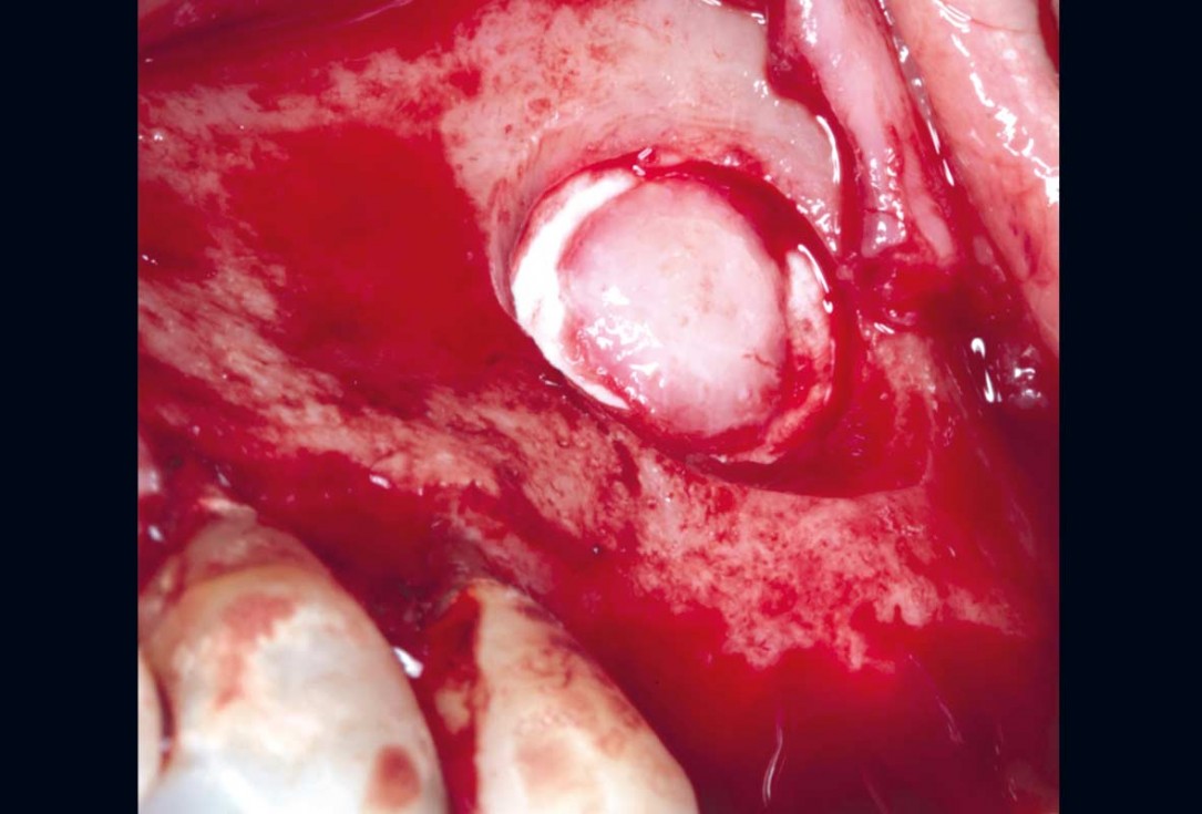
05/35 - Detached sinus membraneMaxillary sinus cyst removal using the Crocodile Technique and subsequent lateral sinus lift - Dres. C. Scognamiglio and A. Perucchi
-
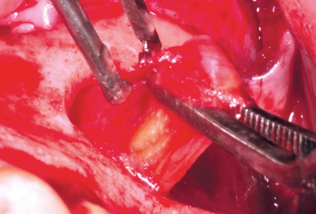
06/35 - Aspiration of the cystic contentsMaxillary sinus cyst removal using the Crocodile Technique and subsequent lateral sinus lift - Dres. C. Scognamiglio and A. Perucchi
-
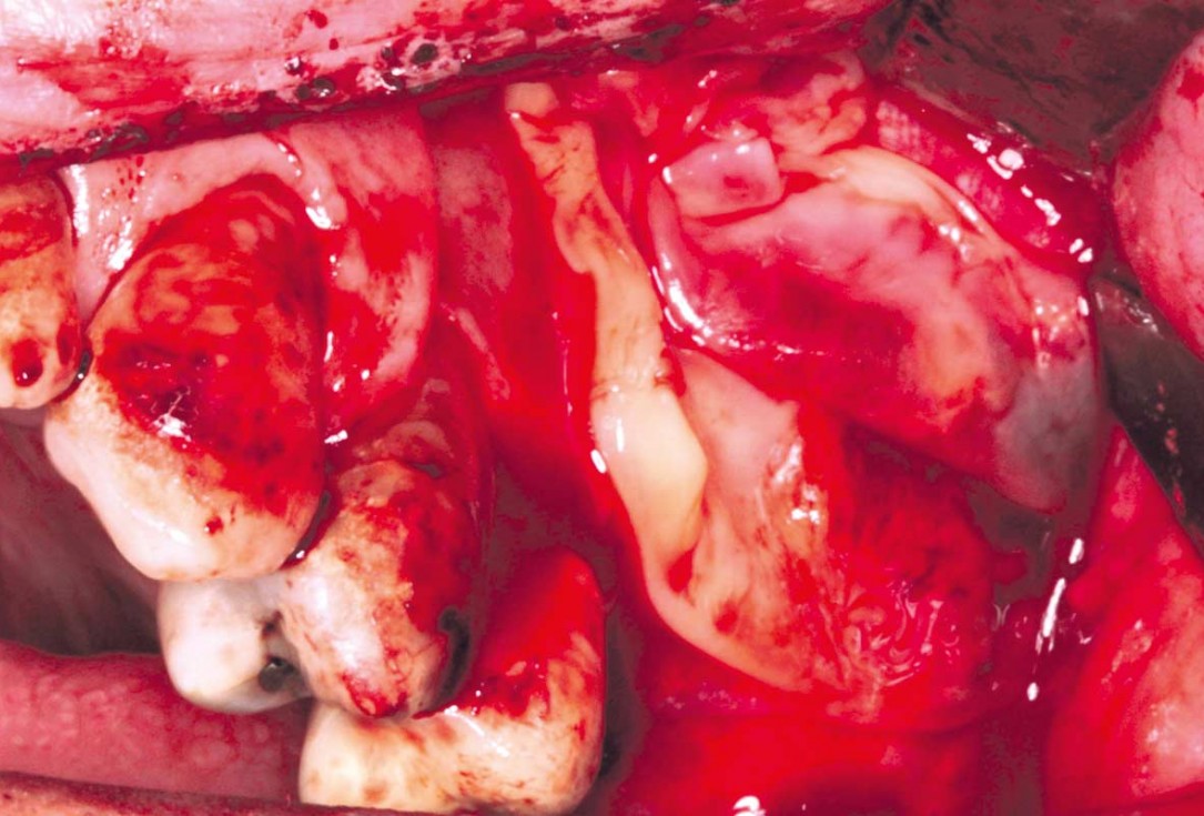
07/35 - Removal of the cyst from the sinusMaxillary sinus cyst removal using the Crocodile Technique and subsequent lateral sinus lift - Dres. C. Scognamiglio and A. Perucchi
-
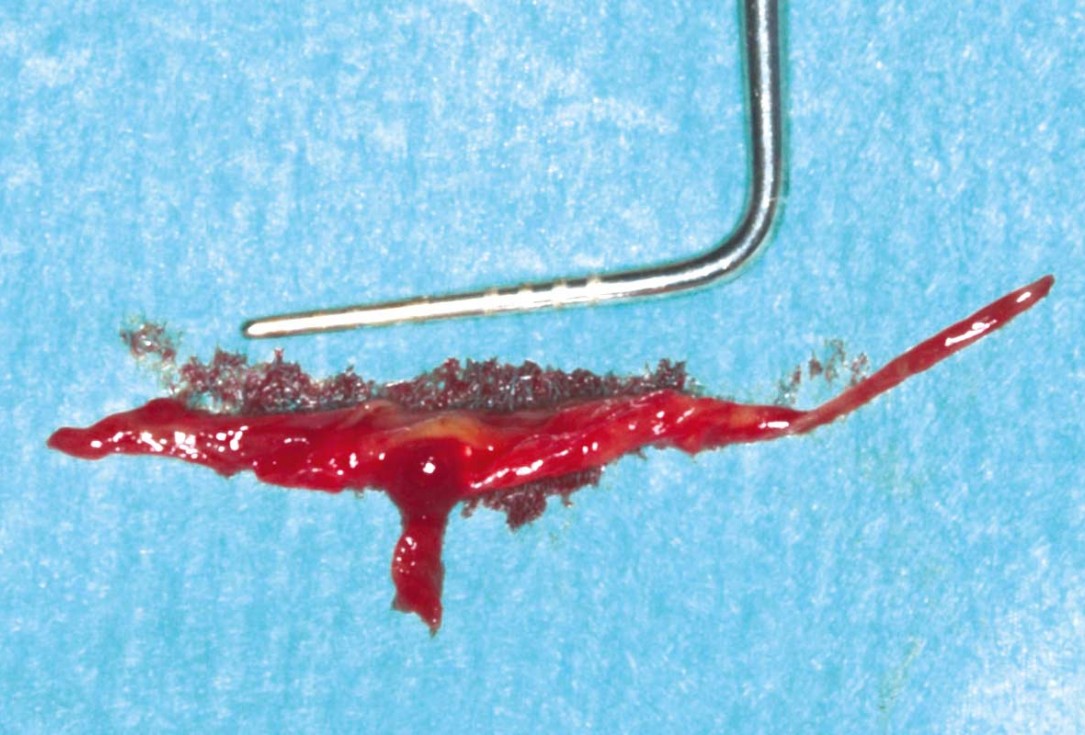
08/35 - CystMaxillary sinus cyst removal using the Crocodile Technique and subsequent lateral sinus lift - Dres. C. Scognamiglio and A. Perucchi
-
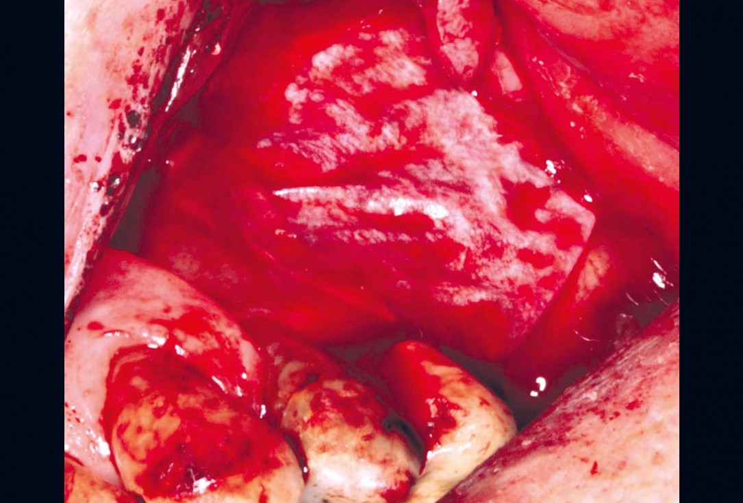
09/35 - Suture of the sinus membraneMaxillary sinus cyst removal using the Crocodile Technique and subsequent lateral sinus lift - Dres. C. Scognamiglio and A. Perucchi
-
10/35 - Protection of the perforation with Jason® membraneMaxillary sinus cyst removal using the Crocodile Technique and subsequent lateral sinus lift - Dres. C. Scognamiglio and A. Perucchi
-
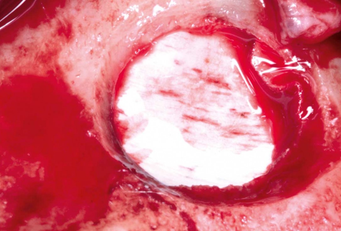
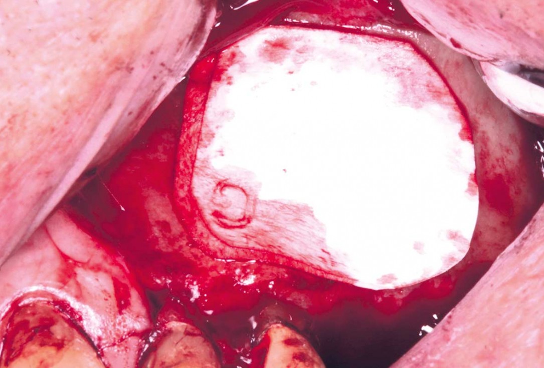
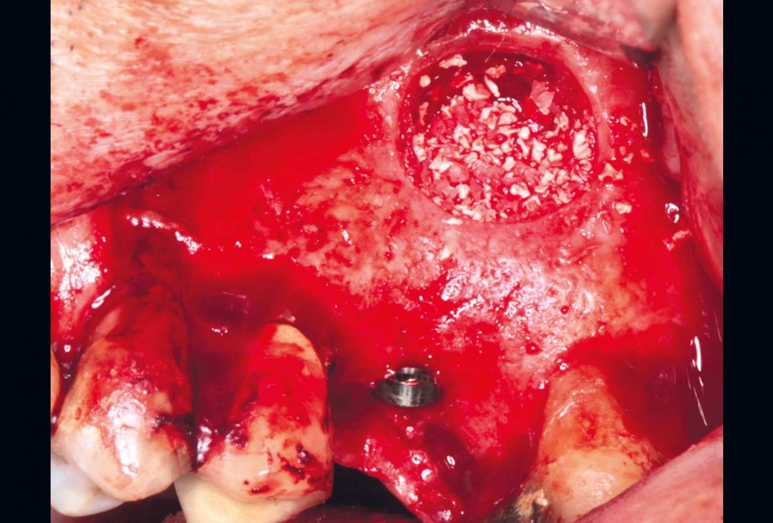
11/35 - Re-Application of the bone window removed at the startMaxillary sinus cyst removal using the Crocodile Technique and subsequent lateral sinus lift - Dres. C. Scognamiglio and A. Perucchi
-
12/35 - Protection of the sinus window with Jason® membrane and L-PRFMaxillary sinus cyst removal using the Crocodile Technique and subsequent lateral sinus lift - Dres. C. Scognamiglio and A. Perucchi
-
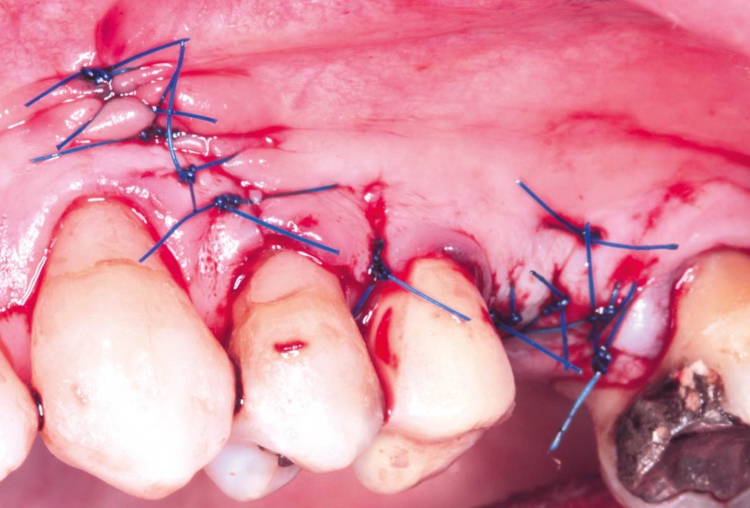
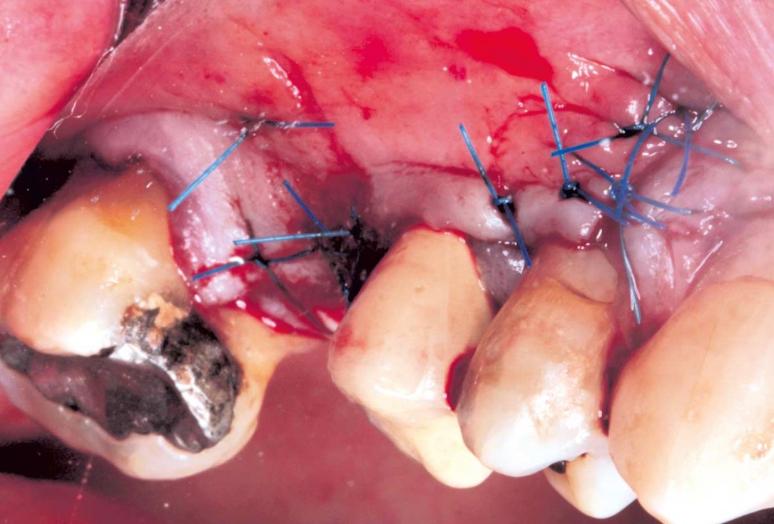
13/35 - SuturesMaxillary sinus cyst removal using the Crocodile Technique and subsequent lateral sinus lift - Dres. C. Scognamiglio and A. Perucchi
-
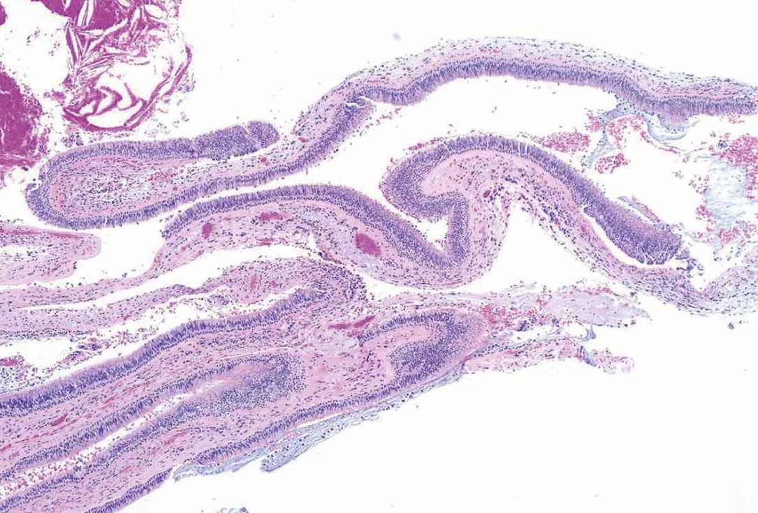
14/35 - Histological analysis shows a salivary retention cystMaxillary sinus cyst removal using the Crocodile Technique and subsequent lateral sinus lift - Dres. C. Scognamiglio and A. Perucchi
-
15/35 - Buccal healing after 4 monthsMaxillary sinus cyst removal using the Crocodile Technique and subsequent lateral sinus lift - Dres. C. Scognamiglio and A. Perucchi
-
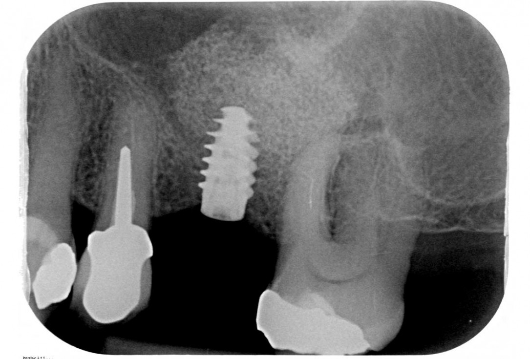
16/35 - X-ray after 5 monthsMaxillary sinus cyst removal using the Crocodile Technique and subsequent lateral sinus lift - Dres. C. Scognamiglio and A. Perucchi
-
17/35 - CBCT after 6 monthsMaxillary sinus cyst removal using the Crocodile Technique and subsequent lateral sinus lift - Dres. C. Scognamiglio and A. Perucchi
-
18/35 - Re-opening of the flap for lateral sinus lift and implant positioningMaxillary sinus cyst removal using the Crocodile Technique and subsequent lateral sinus lift - Dres. C. Scognamiglio and A. Perucchi
-
19/35 - Preparation of the sinus window with piezosurgeryMaxillary sinus cyst removal using the Crocodile Technique and subsequent lateral sinus lift - Dres. C. Scognamiglio and A. Perucchi
-
20/35 - Detachment of the membrane using classic instrumentsMaxillary sinus cyst removal using the Crocodile Technique and subsequent lateral sinus lift - Dres. C. Scognamiglio and A. Perucchi
-
21/35 - Bone augmentation using cerabone®, maxgraft®, and autologous boneMaxillary sinus cyst removal using the Crocodile Technique and subsequent lateral sinus lift - Dres. C. Scognamiglio and A. Perucchi
-
22/35 - Application of Jason® membrane to protect the sinus graftMaxillary sinus cyst removal using the Crocodile Technique and subsequent lateral sinus lift - Dres. C. Scognamiglio and A. Perucchi
-
23/35 - Protection of the sinus window using L-PRFMaxillary sinus cyst removal using the Crocodile Technique and subsequent lateral sinus lift - Dres. C. Scognamiglio and A. Perucchi
-
24/35 - SuturesMaxillary sinus cyst removal using the Crocodile Technique and subsequent lateral sinus lift - Dres. C. Scognamiglio and A. Perucchi
-
25/35 - Post-operative x-rayMaxillary sinus cyst removal using the Crocodile Technique and subsequent lateral sinus lift - Dres. C. Scognamiglio and A. Perucchi
-
26/35 - Radiological follow-up at 6 monthsMaxillary sinus cyst removal using the Crocodile Technique and subsequent lateral sinus lift - Dres. C. Scognamiglio and A. Perucchi
-
27/35 - Mini-invasive second stage, in order to save the keratinized mucosaMaxillary sinus cyst removal using the Crocodile Technique and subsequent lateral sinus lift - Dres. C. Scognamiglio and A. Perucchi
-
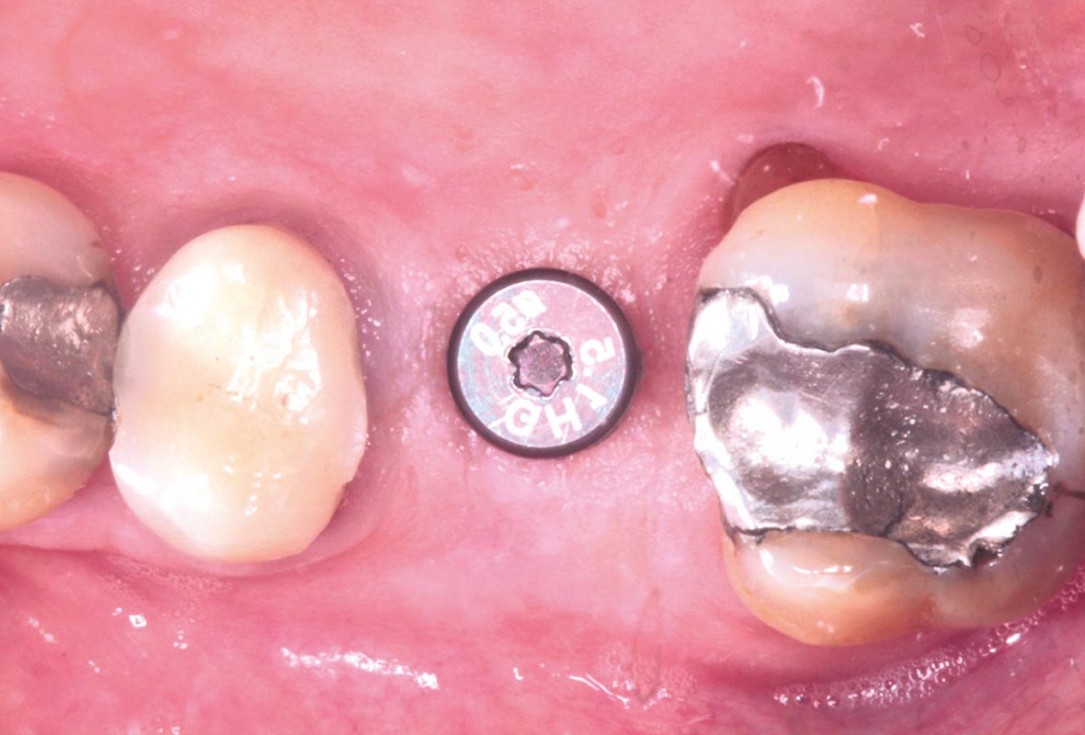
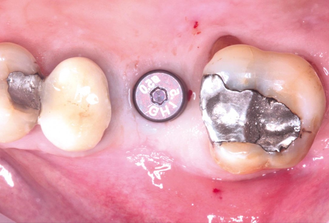
28/35 - Application of the healing cap after the second stageMaxillary sinus cyst removal using the Crocodile Technique and subsequent lateral sinus lift - Dres. C. Scognamiglio and A. Perucchi
-
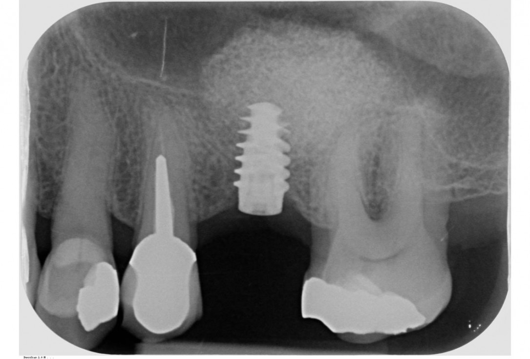
29/35 - X-ray control after the second stageMaxillary sinus cyst removal using the Crocodile Technique and subsequent lateral sinus lift - Dres. C. Scognamiglio and A. Perucchi
-
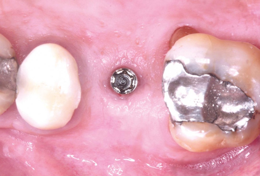
30/35 - Peri-implant tissues at 3 weeks from the second stageMaxillary sinus cyst removal using the Crocodile Technique and subsequent lateral sinus lift - Dres. C. Scognamiglio and A. Perucchi
-
31/35 - Peri-implant tissues at 3 weeks from the second stageMaxillary sinus cyst removal using the Crocodile Technique and subsequent lateral sinus lift - Dres. C. Scognamiglio and A. Perucchi
-
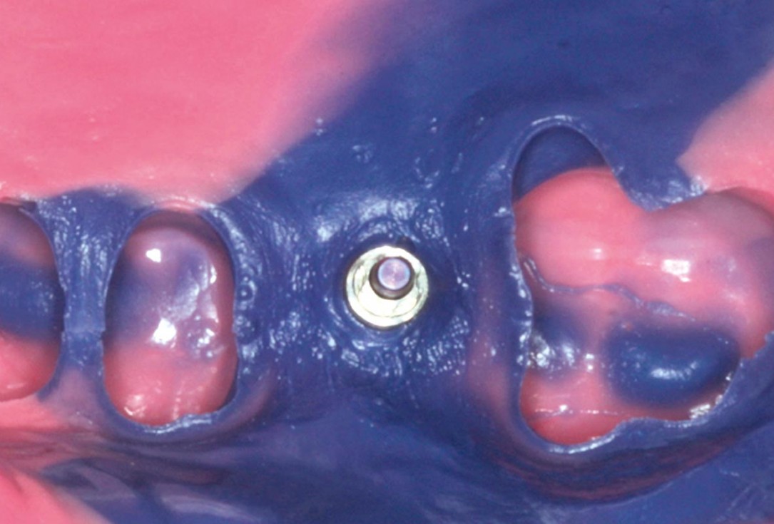
32/35 - Impression with PermadyneMaxillary sinus cyst removal using the Crocodile Technique and subsequent lateral sinus lift - Dres. C. Scognamiglio and A. Perucchi
-
33/35 - Fitting of the zirconia crownMaxillary sinus cyst removal using the Crocodile Technique and subsequent lateral sinus lift - Dres. C. Scognamiglio and A. Perucchi
-
34/35 - Anatomical occlusal filling of the screw channelMaxillary sinus cyst removal using the Crocodile Technique and subsequent lateral sinus lift - Dres. C. Scognamiglio and A. Perucchi
-
35/35 - X-ray follow-up at 8 months from the delivery of the crownMaxillary sinus cyst removal using the Crocodile Technique and subsequent lateral sinus lift - Dres. C. Scognamiglio and A. Perucchi

Preoperative Ortopantomogram of the teeth planned for extraction

DVT image demonstrating horizontal and vertical amount of bone available

Extraction of tooth 21 after endodontic treatment

Implant insertion in atrophic alveolar ridge

Initial clinical situation with broken bridge abutment in regio 12 and tooth 21 not worth preserving

Initial clinical situation with gum recession and labial bone loss eight weeks following tooth extraction

Situation after tooth extraction.

Initial clinical situation

Initial situation after extraction of tooth 21 after 6 months

Situation after tooth removal.

Bone defect in area 11-21 due to two lost implants (periimplantitis) after 15 years of function

Pre-operative clinical situation: severe atrophy of the maxillary bone

60-year-old female patient presented with a chronic infection on tooth #11.
Since she has a high lip line matching the gingival margins of the adjacent central incisor and creating a root eminence is extremely important. For these reasons, the treatment of choice was an allograft bone ring enabling immediate placement of the dental implant with simultaneous regeneration of her ridge.

Initial clinical situation: 9 mm pocket depth associated with root fracture

Initial presentation of failing post retained crown with previous history of failed apicectomies and amalgam tattooing and scar tissue

Initial clinical situation with pronounced vertical and horizontal bone defect

Clinical view 8 weeks after extraction of teeth 25 and 26

Pre-operative: loss of interdental papilla between 12 and 11 associated with gingival inflammation and pus

Preoperative x-ray, multiple residual cysts of the upper jaw

Clinical situation at baseline: Situation after tooth extraction UR1 due to a failed endodontic treatment 3 months previously

Situation before extraction of the teeth

Atrophic alveolar ridge in the left mandible

Instable bridge situation with abscess formation at tooth #15 after apicoectomy

Preoperative clinical situation

Pre-operative radiographic view.

Initial situation pre-op: Central incisors with mobility 3

Initial clinical situation.

Initial situation: X-ray scan reveals eggshell thin sinus floor (1-3 mm) on both sites of the maxilla; green areas indicate the planned maxgraft® bonerings and red areas the planned implants

Pre-surgical situation. Teeth 26 and 27 missing.

Pre-op picture of affected teeth 11 and 21

Preoperative radiological situation

Clinical view of the case.

Preoperative situation – Maxillary defect in area 14-16 (loss of implant 16 due to periimplantitis, tooth 14 extracted recently and area 15 already edentulous for a while)

Initial x-ray showing bone loss around implants placed 5 years ago in another dental clinic

Initial clinical situation

Initial situation - endodontically failing tooth 22, very thin biotype, high lip line and esthetic expectations

Loss of teeth in anterior maxilla caused by periodontitis

Initial situation after root channel treatment

Preoperative CBCT analysis

Preparation of a single tooth defect with severely resorbed vestibular wall

Three implants placed in a narrow posterior mandible

Initial clinical situation with single tooth gap in regio 21

Clinical situation: 71-old patient with atrial fibrillation and Warfarin medication

Initial situation 57-year old female patient. X-ray scan reveals severe bone loss due to inflammation in region 13. Treatment plan was extraction of teeth 13 and 14 and augmentation after healing.

Initial clinical situation, regio #16

Model of the initial defect computed from a CBCT scan - buccal view

Clinical situation

Lateral view of the defect in the posterior right maxilla.

Initial clinical situation.

The patient presented with pathologic mobility of upper left central incisor. Radiographic examination revealed significant circumferential attachment loss with an unfavorable crown to root ratio.

Initial situation – Treatment plan: Replace the adhesive upper left central incisor bridge with a dental implant

Preoperative CBCT: vertical bone defects in the 3rd & 4th quadrant

Initial clinical situation: Free end situation in quadrant three and four

Initial x-ray, tooth 25 compromised and to be extracted

Preoperative x-ray, severe bone atrophy

The patient presented with severe pain in the lateral incisor and a deficient adhesive provisional. Bruxism resulted in canine loss and premature contact in the lateral incisor.

Initial x-ray, ten years post implantationem alio loco, large peri-implant bone loss

