Socket preservation - delayed implantation

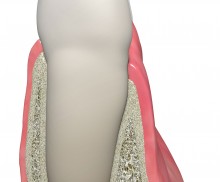
  • Delayed implantation
    Tooth planned for extraction
  • Delayed implantation
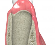
    Fresh extraction socket
  • Delayed implantation
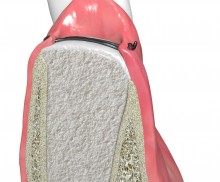
    Filling with maxgraft® and covering with collagen fleece
  • Delayed implantation
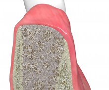
    Healing of the socket after ~3 months
  • Delayed implantation
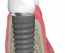
    Implant placed in the healed socket
The alveolar socket is lined with bundle bone—cancellous bone, into which the periodontal fibers infiltrate. This bone is supplied with nutrients by the periodontium and, therefore, resorbs after tooth extraction; in the case of a thin buccal bone, the whole buccal wall may resorb. Socket filling with a bone graft material may not prevent resorption of the bundle bone, but surely helps maintain the volume of the alveoli, thus enabling a later stable implant placement and improving the aesthetic outcome of the final prosthesis. If the implantation is scheduled within the 16 weeks following extraction (delayed implantation), then a grafting material, such as maxgraft® allogenic granules, may be used to support a fast osseous regeneration of the alveoli.
Socket preservation with permamem® - Dr. P. Papi

Pre-operative situation. Lateral view.

Delayed implant placement with GBR and soft tissue augmentation at the aesthetic area  -  2 years follow up – Dr. H. Maghaireh & Dr. V. Ivancheva

Initial situation - broken and missing upper right central incisor (UR1). This tooth was removed long time ago and there were signs of bone loss and resorption due to the bone remodelling. Patient was also undergoing orthodontic treatment due to the loss of mesio-distal space.

Particle application

During application, avoid compressing excessively the particles; less packed particles leave more space for the ingrowth of blood vessels and formation of new bone matrix.

Particle rehydration

Rehydration of maxgraft® granules (as well as cerabone® or maxresorb®) in defect-site blood or saline solution is not necessary, but, because wetted particles tend to stick together, rehydration simplifies both handling and application.

Socket covering

If the bony walls are intact, the application of a barrier membrane is not necessary; however, socket covering is recommended when working with grafting materials. Jason® fleece helps prevent the migration of particles and protects the socket.

Socket with intact walls

Grafting materials act as an osteoconductive scaffold for the formation of new bone within the socket. An ideal grafting material resorbs at the same rate as that of host bone formation; this ensures the volume stability and prevents the ingrowth of soft tissue without causing a delay of the healing process. Due to the presence of preserved collagen, the allogenic maxgraft® granules are characterized by a very high regenerative potential and are completely remodeled into the patient’s own bone within a time period of only ~3–4 months. Therefore, allogenic granules are the best choice for a delayed implantation. Bovine bone granules (cerabone®) or synthetic particles (maxresorb®, collacone® max) may also be used, but generally do not show complete osseous regeneration at 16 weeks post extraction.

"Socket preservation" typically refers to the filling of a socket with intact bony walls. In this case, the application of a membrane is not necessary, but is frequently performed to prevent migration of bone graft particles into the oral cavity. With the purpose of socket covering, the Jason® fleece represents a more cost-effective alternative to barrier membranes. The collagen fleece protects the socket and grafting material, supports wound healing, and may be left exposed for open healing. Another valid solution is to close the socket with mucoderm®. This native three-dimensional collagen matrix helps maintain the soft tissue contours and, specifically for restorations in the front tooth area, ensures optimal aesthetic results. Alternativley, grafted sockts can be covered with a dense PTFE membrane like permamem® in an open healing procedure. Due to its dense structure permamem® acts as an efficient barrier against bacterial and cellular penetration, and may therefore be left in place for open healing in socket or ridge preservation.


Please Contact us for Literature.