Enamel matrix derivative in conjunction with bone grafts

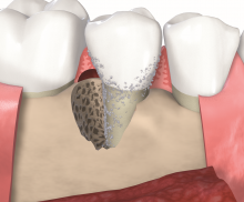
  • Non-self contained intrabony defect.
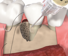
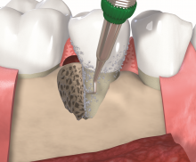
  • Granulation tissue removal and scaling and root planing.
  • Application of Straumann® Emdogain® to the root surface in apico-coronal direction and placement of sutures.
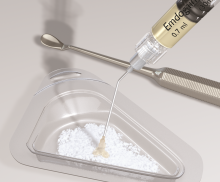
  • Straumann® Emdogain® mixed with particulate bone grafting material.
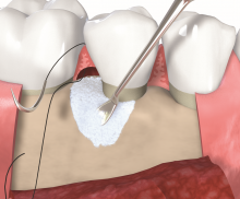
  • Application of the bone graft pre-mixed with Straumann® Emdogain®.
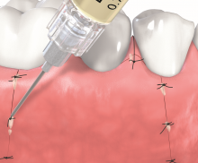
  • Repositioning of the flap and suturing. Remaining Straumann® Emdogain® is applied on the wound margins.
In terms of clinical attachment level gain and probing pocket depth (PPD) reduction, a surgical approach has been proven beneficial for the treatment of deep periodontal intrabony pockets [1, 2, 3]. Enamel matrix derivative (EMD, Straumann® Emdogain®) has been shown to be effective in sites of > 6 mm PPD associated with a radiographic vertical bone loss > 3 mm and to induce the de novo formation of root cementum, periodontal ligament and alveolar bone as evidenced by human histological data [4, 5]. In situations with wide contained or non-contained defects, EMD can be used in conjunction with particulate bone grafts. The graft acts as a scaffold for osteogenic cells, provides space for the regenerative process and supports the soft tissue.

Mixing with bone grafting material

When used in conjunction with bone grafting material, it is recommended to mix Straumann® Emdogain® with the bone graft particles to obtain a paste-like mixture. Also apply Straumann® Emdogain® to the root surface prior to the application of the bone graft/Straumann® Emdogain® mixture.

Barrier membrane

A barrier membrane is not necessarily needed when Straumann® Emdogain® is used. There is currently no evidence that an additional use of a barrier membrane does improve the clinical outcome [6].

Wound healing

Straumann® Emdogain® is indicated to support early soft tissue healing of surgical wounds as part of oral surgical procedures (e.g. flap surgeries, implantation procedures). To improve the soft tissue wound healing after a regenerative periodontal therapy with Straumann® Emdogain®, apply remaining gel on top of the augmentate before final wound closure as well as on the wound margins after suturing.

Application of bone grafting material

Fill the osseous defect as completely as possible with the bone graft granules pre-mixed with Straumann® Emdogain®. Avoid too much compression of the mixture when applied to the defect.

Intrabony defect treated with cerabone® and Straumann® Emdogain® - Dr. D. B. Hangyási

Non-contained intrabony defects (2- or 1-wall defects) usually require volume-providing grafting material in order to prevent flap collapse and to support the soft tissue. Following flap elevation and granulation tissue removal, the root surfaces are freed from plaque and calculus. The remaining smear layer is removed by applying Straumann® PrefGel® for 2 minutes. After thorough rinsing, Straumann® Emdogain® is applied onto the root surfaceto to fully cover the exposed root. The osseous defect is filled with bone substitute particles that were pre-mixed with Straumann® Emdogain®. To support soft tissue wound healing, a final layer of the gel is applied on top of the bone graft before final wound closure.

Different kind of bone substitute materials may be used to fill the defect. cerabone® is a natural bovine bone graft consisting of pure bone mineral. Due to its natural bone structure, cerabone® provides optimal support for bone forming cells and blood vessels. A further alternative are the the allogenic maxgraft® granules or the synthetic material maxresorb®maxresorb® is a biphasic material composed of 60% hydroxyapatite and 40% beta-tricalcium phosphate. While the fast resorption of beta-TCP quickly offers space for new bone formation, the HA component provides volume stability for an extended time period.
 

[1] Badersten et al. J Clin Periodontol. 1981 Feb;8(1):57-72
[2] Badersten et al. J Clin Periodontol. 1985 Jul;12(6):432-40
[3] Heitz-Mayfield et al. J Clin Periodontol. 2002;29 Suppl 3:92-102; discussion 160-2
[4] Sculean et al. J Periodontal Res. 1999 Aug;34(6):310-22
[5] Heijl J Clin Periodontol. 1997 Sep;24(9 Pt 2):693-6
[6] Sculean et al. J Clin Periodontol. 2008 Sep;35(9):817-24