
maxresorb®
- Socket and ridge preservation
- Ridge augmentation
- Intrabony defects (1- to 3-wall)
- Furcation defects (class I-II)
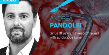
- Sinus floor elevation
- Osseous defects
|

Initial Orthopantomograph X-Ray

Pre-operative x-ray

X-ray control before tooth extraction

DVT image demonstrating horizontal and vertical amount of bone available

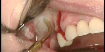
Surgical presentation of the alveolar ridge with reduced amount of horizontal bone available

DVT control after sinusitis surgery, residual bone height 1 mm

Clinical situation before extraction

DVT control after sinusitis surgery, residual bone height 1 mm

DVT image showing the reduced amount of bone available in the area of the mental foramen

X-ray shows a 3-dimensional periondontal defect

Initial situation: Inflammated tooth #12
- Synthetic, resorbable, safe and sterile
- Volume and mechanical graft stability
- Unique multistep production process
- Macropores from 200 to 800 μm
- 60% HA / 40% ß-TCP
- Osteoconductive
- Ultra high interconnected porosity
- Micropores from 1 to 10 μm
Art.-No. | Particle size | Content | ||
|---|---|---|---|---|
20005 | 0.5-1.0 mm (S) | 1x0.5 cc | ||
20010 | 0.5-1.0 mm (S) | 1x1.0 cc | ||
20105 | 0.8-1.5 mm (L) | 1x0.5 cc | ||
20120 | 0.8-1.5 mm (L) | 1x2.0 cc |
Art.-No. | Dimensions | Content | ||
|---|---|---|---|---|
21211 | 20x10x10 mm | 1xblock | ||
21221 | 20x20x10 mm | 1xblock | ||

The specific composition of maxresorb® promotes the fast formation of new vital bone, ensuring a long-term mechanical and volume stability. The osteoconductivity of maxresorb® is achieved by a matrix of interconnecting pores (with a size ranging between 200 and 800 μm) and a very high porosity of approx. 80%. The high microporosity and nano-structured surface facilitate the uptake and adsorption of blood, proteins, and stem cells. The macropores are ideal for the ingrowth of osteogenic cells and the bony integration.
Please find our free webinars at www.botiss-webinars.com
Kostenfreie Webinare zu Schulungszwecken finden Sie unter www.botiss-webinars.com
Please find our free webinars at www.botiss-webinars.com
Please find our free webinars at www.botiss-webinars.com
Please find our free webinars at www.botiss-webinars.com
Please find our free webinars at www.botiss-webinars.com
Please find our free webinars at www.botiss-webinars.com


