maxgraft® bonering - one-stage bone augmentation and immediate implant placement

maxgraft® bonering

One-stage bone augmentation and implantation
maxgraft® bonering is a prefabricated ring of processed allogenic donor bone, which is placed press-fit into a trephine drill-prepared ring bed. At the same time, an implant is inserted into the ring. The bony integration of both maxgraft® bonering and the implant occurs via the surrounding vital bone. The maxgraft® bonering technique allows bone augmentation and implantation in a one-stage procedure. The technique is suitable for virtually all indications, including sinus lift with limited maxillary bone height.
  • Vertical augmentation (in combination with horizontal augmentation)
  • Single tooth gap
  • Edentulous space
  • Sinus lift

Wound closure

Carefully consider the soft tissue situation prior to surgical intervention! Please keep in mind that a tension-free closure of the flap is essential for the success of the procedure.

Rehydration

The processing of maxgraft® products preserves the natural collagen content of the bone tissue and a residual water content of <10%. Thus, maxgraft® bonering does not need to be rehydrated, but it is recommended. Rehydration enhances the flexibility of the ring, therefore it is less prone to crumble or break and can be more easily adapted to the defect area. Above that, preparation of the ring bed using the maxgraft® bonering surgical kit ensures close contact of the bone ring to the vital bleeding bone, leading to the supply of blood to the ring. Thus, enabling fast bony integration of implant and bone graft.

Re-entry

Once maxgraft® bonering is fixed with a suitable implant, it provides great primary stability. Implant loading should be performed 6 months after transplantation to avoid resorption of the bone graft caused by a lack of mechanical stimulus.

Combination with cerabone®

The combination of maxgraft® bonering with a xenogenic material (cerabone®) combines the advantages of both materials; the biological potential of maxgraft® bonering induces fast incorporation of graft and implant, the volume-stable cerabone® acts as a barrier against resorption and improves the esthetic outcome.

Graft exposure

Wound dehiscence and graft exposure can be common complications of augmentation. After removal of necrotic soft tissue and infected hard tissue (use rotating instruments if necessary) the augmented area should be rinsed with chlorhexidine. Subsequently, the graft has to be covered again, if necessary, by harvesting a palatal soft tissue transplant.

Vertical augmentation and sinus lift with maxgraft® bonering - Dr. O. Yüksel and Dr. B. Giesenhagen

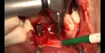
Vertical augmentation: Preparation of ring bed in atrophic mandibula (third quadrant)

Restoration of buccal layer with maxgraft® bonering - A. Patel

Initial situation: missing incisor with loss of buccal wall

Guided bone ring procedure in aesthetic zone - Dr. K. Chmielewski & Dr. O. Yükse

Planning the surgery with CoDiagnostix® for Straumann® Guided Surgery

Sinus Floor Elevation with maxgraft® bonering - Dr. B. Giesenhagen

X-ray scan reveals initial situation with maxillary bone height in regio 15 of 1.5 mm

Restoration of buccal bone lamella in aesthetic zone with maxgraft® bonering - Dr. A. Patel

Initial situation: bone loss due to lack of physical load of bridge retained region 11

Restoration of all four incisors with two maxgraft® bonering - Dr. B Giesenhagen

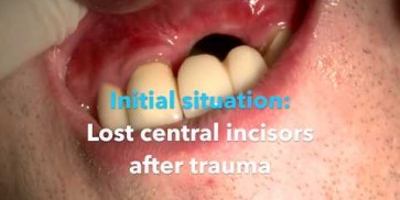
Initial situation pre-op: Central incisors with mobility 3

Periimplantitis treatment with maxgraft® bonering - Dr. B. Giesenhagen

Severe periimplantitis at tooth 15 with bone loss up to 1/3 of the implant

Sinus Floor Elevation with maxgraft® bonering and subcrestal implantation in an eggshell thin sinus - Dr. K. Chmielewski

Initial situation: X-ray scan reveals eggshell thin sinus floor (1-3 mm) on both sites of the maxilla; green areas indicate the planned maxgraft® bonerings and red areas the planned implants

Advanced vertical augmentation in posterior maxilla with maxgraft® bonering - Dr. A. Isser

Initial situation 57-year old female patient. X-ray scan reveals severe bone loss due to inflammation in region 13. Treatment plan was extraction of teeth 13 and 14 and augmentation after healing.

Augmentation of buccal and lingual bone lamella with maxgraft® bonering - Dr. B. Giesenhagen

X-ray scan: initial situation loss of two wall bony defect with loss of buccal and lingual lamella
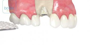
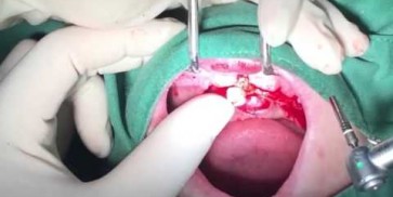
Bone augmentation in the aesthetic zone with maxgraft® bonering – Dr. R. Cutts

Initial presentation of failing post retained crown with previous history of failed apicectomies and amalgam tattooing and scar tissue

  • Ring-shaped spongiosa block from living donors
  • Simultaneous implant placement and bone augmentation
  • Significant reduction of treatment time
  • Fast incorporation and complete remodeling to patients own bone
  • No second surgical procedure necessary
  • Storable at room temperature for 5 years

Art.-No.

Dimensions

Content

33160

height 10 mm, diameter 6.0 mm/3.3 mm (outer/inner diameter)

1x cancellous ring

33170

height 10 mm, diameter 7.0 mm/3.3 mm (outer/inner diameter)

1x cancellous ring

33174

height 10 mm, diameter 7.0 mm/4.1 mm (outer/inner diameter)

1x cancellous ring

Art.-No.

Dimensions

Content

33000

maxgraft® bonering surgical kit

1 set

33010

bonering fix

1 x

maxgraft® bonering technique animation
Live OP vertikale Augmentation mit maxgraft® bonering Dr. A. Vossenberg
Bone Ring Technique surgery performed by Dr. Orcan Yüksel and Dr. Kris Chmielewski
Live-surgery maxgraft® bonering Dr. Bernhard Giesenhagen
Live surgery maxgraft® bonering Dr. Bernhard Giesenhagen
maxgaft® bonering surgery David Furze
Live surgery maxgraft® bonering Dr. Bernhard Giesenhagen & Dr. Orcan Yüksel
Dr. Orcan Yüksel about the maxgraft® bonering
maxgraft® bonering surgery by Dr. Bernhard Giesenhagen
maxgraft® bonering - one-stage bone augmentation and immediate implant placement

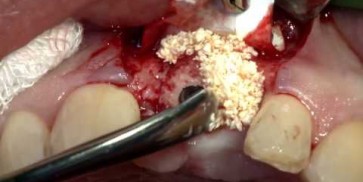
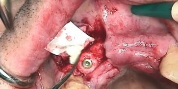
Compared to the classical two-stage augmentation with bone blocks, this technique reduces the entire treatment period by several months and saves the re-entry. maxgraft® bonering is suitable for vertical and horizontal augmentation and promotes new bone formation, therefore simplifying the surgical treatment. With the maxgraft® bonerig surgical kit, botiss biomaterials provides all necessary instruments to apply the maxgraft® bonering technique. The kit includes two convenient sizes of trephines, which precisely match the maxgraft® bonering diameters. The planators allow the paving of the local bone to create a congruent and fresh contact surface of the implant area. The diamond disc and the diamond tulip can be used to shape maxgraft® bonering for an excellent adjustment to the local bone and for an improved soft tissue healing. Altogether, the adaptation of maxgraft® bonering with the instrument kit allows optimal preconditions for the bony ingrowth. All instruments are made of high-quality surgical steel.

[1] Flanagan D. (2016). Cylindrical Ringbone Allograft to Restore Atrophic Implant Sites: A Pilot Study. Journal of Oral Implantology 2016;42(2):159-163.
[2] Miller at el. (2017). Use of the Straumann® AlloGraft Ring With Simultaneous Implant Placement: A Novel Approach. Compendium of Continuing Education in Dentistry Nov/Dec 2017, Vol 38, Issue 11 https://www.aegisdentalnetwork.com/cced/2017/11/use-of-the-straumann-allograft-ring-with-simultaneous-implant-placement-a-novel-approach
[3] Giesenhagen G, Martin N, Donkiewicz P, Kacarević ZP, Smeets R, Jung O, Schnettler R, Barbeck M (2018). Vertical bone augmentation in a single-tooth gap with an allogenic bone ring: Clinical considerations. J Esthet Restor Dent. 2018;1–4. https://doi.org/10.1111/jerd.12392
[4] Benlidayi E, Salimov F, Tukel C, Yüksel O (2018). Comparison of autogenous and allograft bone rings in surgically created vertical bone defects around implants in a sheep model. Clin Oral Implants Res. 2018. https://www.ncbi.nlm.nih.gov/m/pubmed/30281857/
[5] Giesenhagen B, Martin N, Jung O, Barbeck M (2019). Bone Augmentation and Simultaneous Implant Placement with Allogenic Bone Rings and Analysis of Its Purification Success. Materials 2019 9 12, 1291; https://doi.org/10.3390/ma12081291