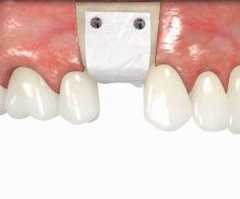
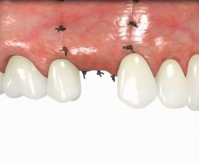
Bone ring technique

  • Bone ring technique
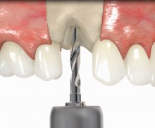
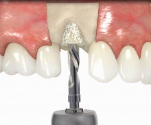
    Pilot drill
  • Bone ring technique
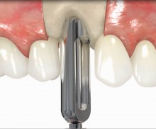
    Ring bed preparation with a trephine
  • Bone ring technique
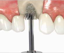
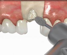
    Ring bed preparation with a planator
  • Bone ring technique
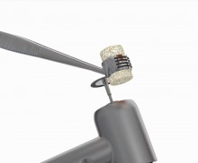
    Ring length adjustment with a diamond disc
  • Bone ring technique
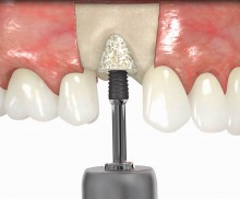
    Implant drill through the inserted bone ring
  • Bone ring technique
    Implant insertion through the bone ring
  • Bone ring technique
    Sharp edge smoothening
  • Bone ring technique
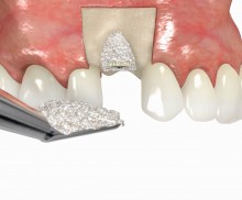
    Covering with cerabone®
  • Bone ring technique
    Covering with pinned Jason® membrane
  • Bone ring technique
    Tension-free wound closure
The bone ring technique is a surgical methodology that allows bone augmentation and implantation in a one-stage procedure. Compared to the more conventional two-stage approach, the main advantage of the bone ring technique is that of significantly reducing the treatment time by several months. The bone ring technique enables vertical/horizontal augmentation and formation of new bone, thereby simplifying the surgical treatment of 3-dimensional bone defects. The technique is applicable for almost all indications, including sinus lift with a limited maxillary bone height. According to the original approach, the augmentation is performed with autologous bone rings, which are harvested intraorally (mostly from the chin area or palate) with a trephine. The bone ring is then transferred to the trephine drill-prepared recipient site. The implant bed is thus prepared in the local bone through the ring opening, and the implant is inserted through the ring. The bony integration of the bone ring and implant occurs via the surrounding bone.
Vertical augmentation and sinus lift with maxgraft® bonering - Dr. O. Yüksel and Dr. B. Giesenhagen

Vertical augmentation: Preparation of ring bed in atrophic mandibula (third quadrant)

Guided bone ring procedure in aesthetic zone - Dr. K. Chmielewski & Dr. O. Yükse

Planning the surgery with CoDiagnostix® for Straumann® Guided Surgery

Periimplantitis treatment with maxgraft® bonering - Dr. B. Giesenhagen

Severe periimplantitis at tooth 15 with bone loss up to 1/3 of the implant

Restoration of buccal bone lamella in aesthetic zone with maxgraft® bonering - Dr. A. Patel

Initial situation: bone loss due to lack of physical load of bridge retained region 11

bone augmentation in the aesthetic zone with maxgraft® bonering -Dr. R. Cutts

Clinical situation: 71-old patient with atrial fibrillation and Warfarin medication

Restoration of all four incisors with two maxgraft® bonering - Dr. B Giesenhagen

Initial situation pre-op: Central incisors with mobility 3

Sinus Floor Elevation with maxgraft® bonering - Dr. B. Giesenhagen

X-ray scan reveals initial situation with maxillary bone height in regio 15 of 1.5 mm

Sinus Floor Elevation with maxgraft® bonering and subcrestal implantation in an eggshell thin sinus - Dr. K. Chmielewski

Initial situation: X-ray scan reveals eggshell thin sinus floor (1-3 mm) on both sites of the maxilla; green areas indicate the planned maxgraft® bonerings and red areas the planned implants

Restoration of buccal layer with maxgraft® bonering - A. Patel

Initial situation: missing incisor with loss of buccal wall

Advanced vertical augmentation in posterior maxilla with maxgraft® bonering - Dr. A. Isser

Initial situation 57-year old female patient. X-ray scan reveals severe bone loss due to inflammation in region 13. Treatment plan was extraction of teeth 13 and 14 and augmentation after healing.

Augmentation of buccal and lingual bone lamella with maxgraft® bonering - Dr. B. Giesenhagen

X-ray scan: initial situation loss of two wall bony defect with loss of buccal and lingual lamella

Bone augmentation in the aesthetic zone with maxgraft® bonering – Dr. R. Cutts

Initial presentation of failing post retained crown with previous history of failed apicectomies and amalgam tattooing and scar tissue

Wound closure

Carefully consider the soft tissue situation prior to surgical intervention! Please keep in mind that a tension-free closure of the flap is essential for the success of the procedure.

Rehydration

The processing of maxgraft® products preserves the natural collagen content of the bone tissue and a residual water content of <10%. Thus, maxgraft® bonering does not need to be rehydrated, but it is recommended. Rehydration enhances the flexibility of the ring, therefore it is less prone to crumble or break and can be more easily adapted to the defect area. Above that, preparation of the ring bed using the maxgraft® bonering surgical kit ensures close contact of the bone ring to the vital bleeding bone, leading to the supply of blood to the ring. Thus, enabling fast bony integration of implant and bone graft.

Re-entry

Once maxgraft® bonering is fixed with a suitable implant, it provides great primary stability. Implant loading should be performed 6 months after transplantation to avoid resorption of the bone graft caused by a lack of mechanical stimulus.

Combination with cerabone®

The combination of maxgraft ® bonering with a xenogenic material (cerabone®) combines the advantages of both materials; the biological potential of maxgraft® bonering induces fast incorporation of graft and implant, the volume-stable cerabone® acts as a barrier against resorption and improves the esthetic outcome.

Graft exposure

Wound dehiscence and graft exposure can be common complications of augmentation. After removal of necrotic soft tissue and infected hard tissue (use rotating instruments if necessary) the augmented area should be rinsed with chlorhexidine. Subsequently, the graft has to be covered again, if necessary, by harvesting a palatal soft tissue transplant.

Augmentation durch Einsetzen eines maxgraft® bonering

botiss offers prefabricated allogenic bone rings, whose use minimizes the risks associated with the harvesting procedure and reduces the post-operative pain as well as the risk of infection and operation time. maxgraft® bonering is a sterile, highly safe allograft product derived from the femoral heads of living human donors. The prefabricated rings can be placed press-fit into the prepared ring bed.

In addition, botiss offers a surgical kit that provides all necessary instruments for the bone ring technique. The first step of the surgical procedure is the determination of the position of the implant using the pilot drill; the ring bed is then prepared with a trephine. Subsequently, the use of a planator enables an even paving of the local bone to achieve an optimal contact with maxgraft® bonering as well as the removal of the cortical layer, which improves the graft revascularization. With maxgraft® bonering, a vertical bone augmentation of 2–3 mm above bone level can be easily performed. For the augmentation of a severely atrophic mandibular, a width of the ridge (in the case of a parallel-walled ridge) of at least 4 mm is necessary for a successful application of maxgraft® bonering. The technique is also applicable for a one-stage sinus lift with minimal maxillary bone height. In this case, the implant is fixed in the sinus with the allogenic bone ring; these are held together with a membrane screw.

It is recommended to fill the residual defect volume with a slowly resorbable bone regeneration material (i.e., cerabone®) and to cover the augmentation site with a collagen membrane. The pericardial Jason® membrane is an ideal solution in such a situation due to its long-term barrier function. A tension-free suturing of the operation site minimizes the risk of tissue perforation and/or graft exposure.

Please Contact us for Literature.