mucoderm® - Greffon de tissus mous en 3D

mucoderm®

Greffon de tissus mous en 3D
mucoderm® est une matrice de collagène naturel de type I/III dérivé du derme porcin ayant subi un processus de purification en plusieurs étapes, qui élimine toute substance immunogène potentielle. La matrice résiduelle est une membrane constituée de collagène et d’élastine. mucoderm® favorise la revascularisation et une intégration rapide des tissus mous, et représente une alternative valable aux greffes autologues de tissu conjonctif.
  • Augmentation des tissus mous
  • Recouvrement des alvéoles d’extraction
  • Récessions gingivales
  • Greffe de tissus mous en association avec une ROG/RTG
  • Élargissement de la gencive attachée

Immobilisation

Il convient d’assurer un contact de mucoderm® avec le lit périosté de la plaie et une immobilisation, en fixant la matrice au périoste par une suture discontinue simple ou une suture croisée.

Éviter les bords coupants

Le fait de découper les bords d’une matrice peu après sa réhydratation évite d’endommager les tissus gingivaux durant la fermeture du lambeau.

Technique de tunnel

Pour la technique de tunnel, un temps de réhydratation prolongé de mucoderm®, de 10 à 20 min, est recommandé. La fixation de la matrice par une suture discontinue simple ou par une suture croisée est requise.

Recouvrement de récessions

L’exposition de la matrice mucoderm® sera toujours évitée lors des procédures de recouvrement de récessions. S’assurer que le lambeau repositionné recouvre complètement la matrice mucoderm®. Le fait de réaliser une fermeture primaire sur la greffe de mucoderm® permet aux vaisseaux sanguins de pénétrer et d’incorporer le matériau de greffe de tissus mous. Une exposition précoce peut entraîner l’échec de la greffe de tissus mous.

Straumann® Emdogain®

Les récessions gingivales peuvent être traitées en utilisant mucoderm® et Straumann® Emdogain® ensemble dans le cadre d’une même approche. Les avantages potentiels d’ajouter Straumann® Emdogain® à une procédure de recouvrement radiculaire par mucoderm® sont décrits à la section « Matrice de collagène et dérivé de la matrice amélaire ». Veiller à toujours appliquer Straumann® Emdogain® sur la totalité des surfaces radiculaires avant de leur appliquer la matrice mucoderm®.

cerabone® and mucoderm® for immediacy in esthetic zone -Dr. M Motta

Initial view of the case. Discoloration of 1.1 and mild class I gingival recession

botiss mucoderm® for treatment of single gingival recession - Clinical case

Initial clinical situation with Miller class 1 recession

Tooth extraction and socket sealing with mucoderm® - Dr. A. Rossi

Initial clinical situation showing strongly compromised tooth 21

Ridge augmentation in the maxilla with maxgraft® bonebuilder in the aesthetic zone - Dr. M. Kristensen

Bone defect in area 11-21 due to two lost implants (periimplantitis) after 15 years of function

Socket seal with mucoderm® punch

Intact socket following atraumatic tooth extraction

Multiple gingival recessions treated by MCAF in conjunction with mucoderm® - Kasaj

Pre-surgical clinical situation. Multiple adjacent gingival recessions at teeth 12,13 and 14.

Multiple gingival recessions treated by MCAT with mucoderm® and Straumann® Emdogain® (2) - Rathe

Pre-operative clinical situation. Shallow multiple adjacent gingival recessions in the first quadrant.

Immediate implantation with maxresorb® - Dr. M. Frosecchi

X-ray control before tooth extraction

botiss maxgraft® bonebuilder and vestibuloplasty with mucoderm® for ridge augmentation - Clinical case

Preoperative situation – Maxillary defect in area 14-16 (loss of implant 16 due to periimplantitis, tooth 14 extracted recently and area 15 already edentulous for a while)

Regeneration with mucoderm® after fibroma resection

Initial clinical situation shows an odontogenic fibroma that was growing for years

cerabone® and mucoderm® for immediate implantation in the aesthetic area - Dr. D. Robles

Initial clinical situation - Central incisors with dental destruction and periapical pathology

Mucosal thickening around bone level implants - Dr. A. Puisys

Full-thickness flap preparation bucally and lingually

GBR and soft tissue augmentation with cerabone® and mucoderm® - H. Maghaireh & V. Ivancheva

Initial situation: missing teeth #11 & 12 and badly broken #21 root

Augmentation of a traumatic soft tissue defect-Rathe

Initial clinical situation with traumatic loss of tooth 21

GBR together with soft tissue augmentation with mucoderm® and maxresorb® - Scherg

Longitudinal fracture on the root resected tooth 21 with visible buccal fistula

Post extraction socket sealing with mucoderm® - Dr. M. Frosecchi

X-ray showing endodontic failure of the molar

Block augmentation with maxgraft® block and mucoderm® - Dr. K. Chmielewski

Initial situation before surgery. Patient lost central incisors 1 month ago due to endodontic failures

Regenerative corticotomy to compensate lower incisor malocclusion with cerabone® and mucoderm®

Initial view of the clinical case: Class III malocclusion
Treatment plan: Regenerative corticotomy (PAOO)

Soft tissue thickening with mucoderm® - Dr. G. de Deus

Initial clinical view of the case. Soft tissue dehiscence around implants 26.

Single gingival recession treated with the envelope technique in conjunction with mucoderm® - Kasaj

Gingival recession at tooth 13. Free gingival graft (FGG) of a previous surgery for root coverage visible.

Widening of the peri-implant keratinized mucosa-Horváth

Lack of sufficient keratinized mucosa following extensive horizontal ridge augmentation

Treatment of a soft tissue deficiency with mucoderm® - Dr. F. Rojas-Vizcaya

Initial clinical situation showing severe soft tissue loss

botiss mucoderm® for coverage of multiple recessions - clinical case by Dr. S. Stavar

Initial clinical situation. Multiple adjacent gingival recessions in regio 11-13.

botiss maxgraft® bonebuilder aesthetic reconstruction - Clinical case

Pre-operative clinical situation: changed color in the gingiva in the front maxilla

botiss-cerabone-permamem-maxgraft-mucoderm-horizontal-gbr-maghaireh

Initial situation - A young female 34 years old lost her front teeth in an surfing accident and she had a 5 unit bridge supported by her upper left lateral and right canine. The restoration failed and both supporting crowns have exposed and leaking margins.

  • Revascularisation et intégration rapides
  • Remplacement des tissus mous sans prélèvement d’un greffon autologue palatin
  • Remodelage complet dans les propres tissus du patient
  • Temps de résorption d’environ 6 à 9 mois
  • Mise en place et fixation aisées
  • Possibilité de découpe pour des procédures spécifiques
  • Épaisseur d’environ 1,2-1,7 mm

Art. -No.

Size

Content

701520

15x20

1 matrix

702030

20x30

1 matrix

703040

30x40

1 matrix

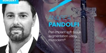
Soft tissue augmentation using mucoderm® (2-year follow up) - live surgery by Dr. A. Pandolfi
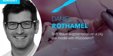
Soft tissue thickening on pig jaw by PD Dr. Dr. D. Rothamel
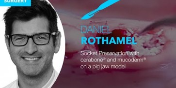
Socket seal on pig jaw by PD Dr. Dr. D. Rothamel
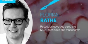
Recession coverage using the MCAT technique and mucoderm® - live surgery by Dr. F. Rathe
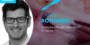
Vestibuloplasty on pig jaw by PD Dr. Dr. D. Rothamel
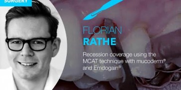
Recession coverage using the MCAT technique in conjunction with mucoderm® and Straumann® Emdogain®- live surgery by Dr. F. Rathe
Corrosion preparation showing vascular network running through the mucoderm® matrix
mucoderm® - 3D soft tissue graft

Après la mise en place, le sang du patient s’infiltre dans la greffe de mucoderm® à travers le réseau tridimensionnel des tissus mous, en amenant des cellules hôtes à la surface de la greffe de tissus mous et en déclenchant le processus de revascularisation. Une revascularisation importante peut débuter après l’implantation, en fonction de l’état de santé du patient. mucoderm® constitue une alternative sûre à la greffe autologue de tissu conjonctif, et convient à une large palette d’indications de greffe de tissus mous.

Please Contact us for Literature.