Horizontal ridge augmentation with maxgraft® cortico - M.Sc. E. Kapogianni
-
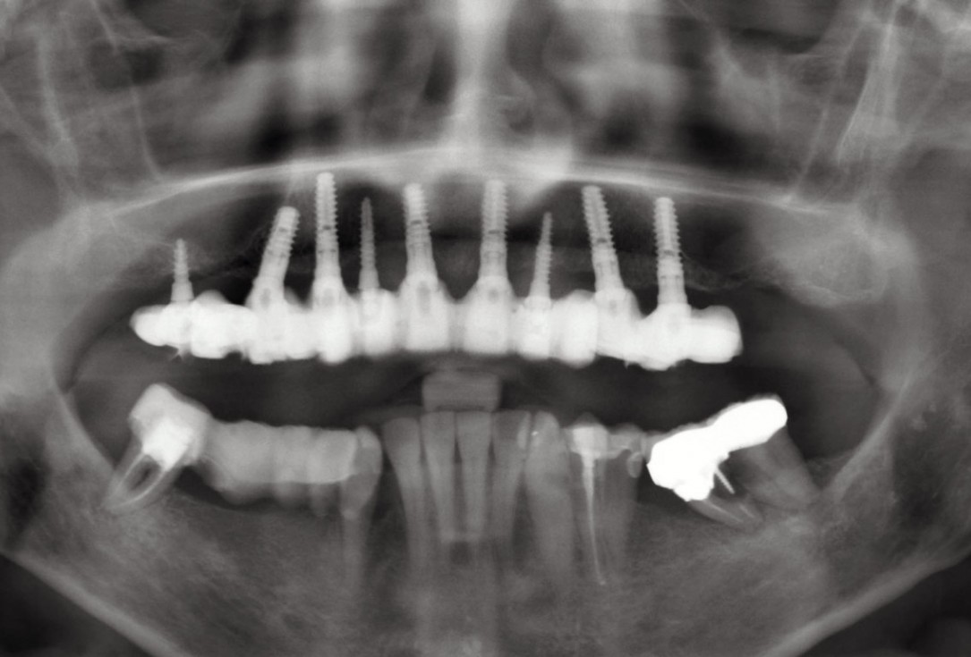
01 / 20 - OPG of the initial situation – provision of missing denture in regio 44 to 47 by a resin-retained bridgeHorizontal ridge augmentation with maxgraft® cortico - M.Sc. E. Kapogianni
-
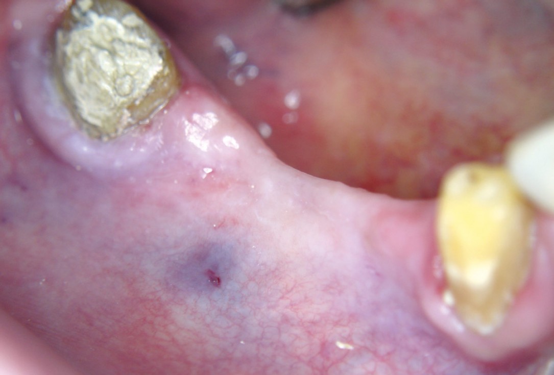
02 / 20 - Thin soft tissue and reduced mandibular widthHorizontal ridge augmentation with maxgraft® cortico - M.Sc. E. Kapogianni
-
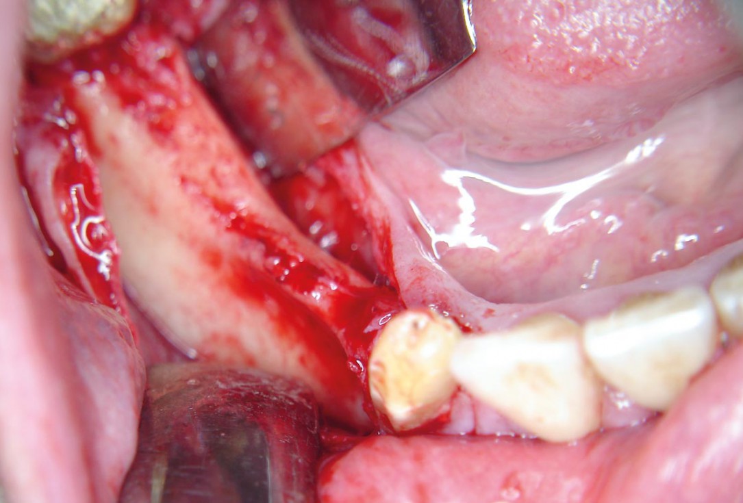
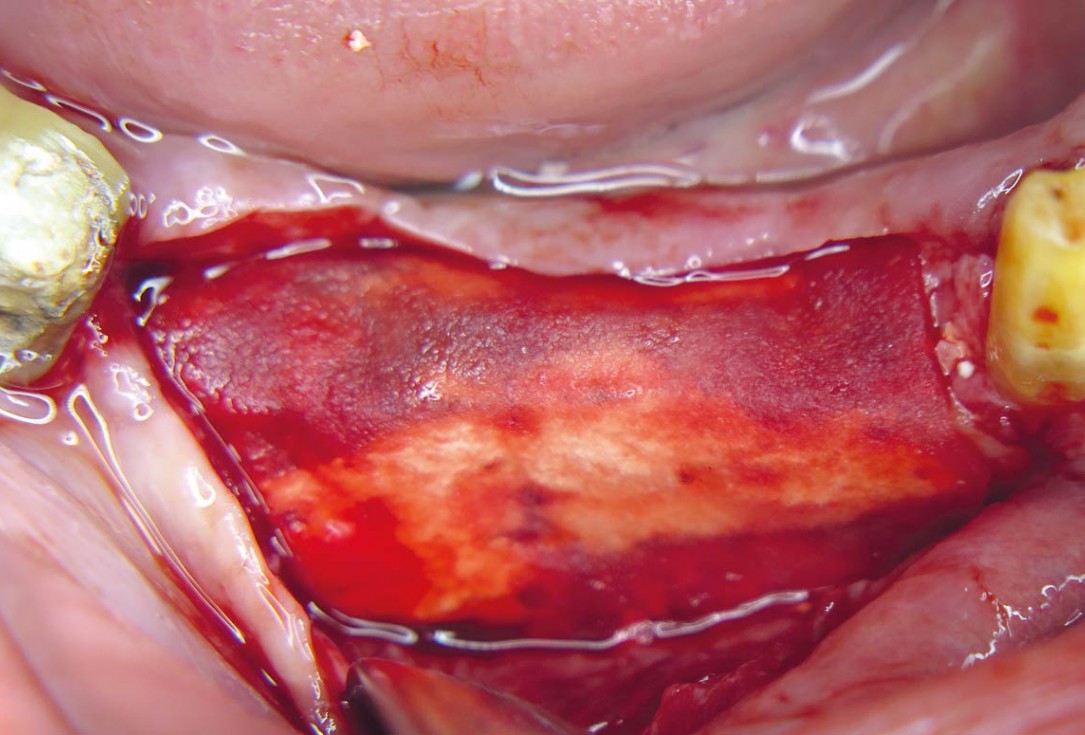
03 / 20 - Flap projection shows pronounced bone lossHorizontal ridge augmentation with maxgraft® cortico - M.Sc. E. Kapogianni
-
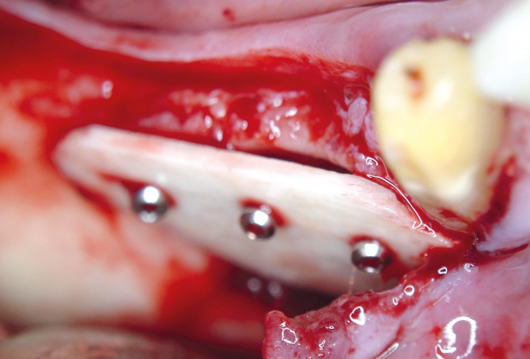
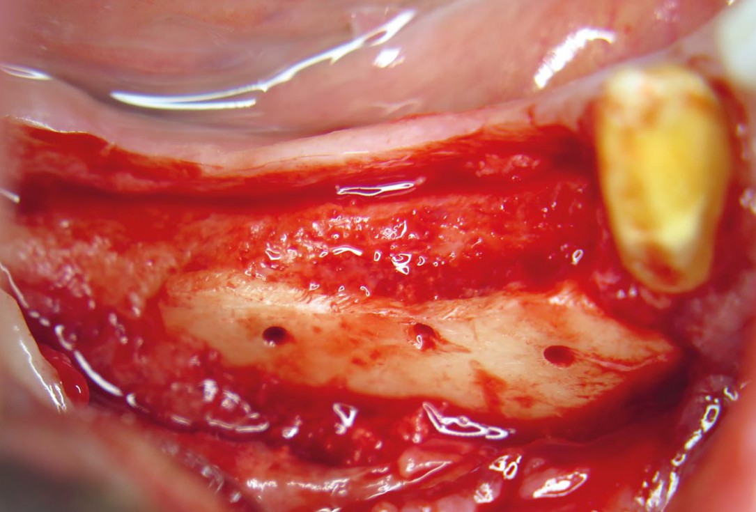
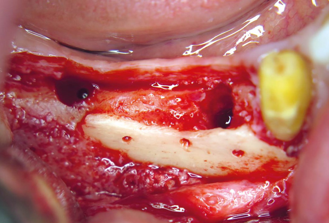
04 / 20 - Decortication of the host bone and fixation of a defect-adapted cortical plateHorizontal ridge augmentation with maxgraft® cortico - M.Sc. E. Kapogianni
-
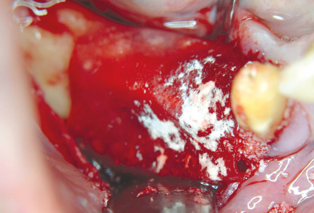
05 / 20 - Defect-filling and contouring with cancellous allogenic bone chipsHorizontal ridge augmentation with maxgraft® cortico - M.Sc. E. Kapogianni
-
06 / 20 - Augmentation site covering by application of a collprotect® membraneHorizontal ridge augmentation with maxgraft® cortico - M.Sc. E. Kapogianni
-
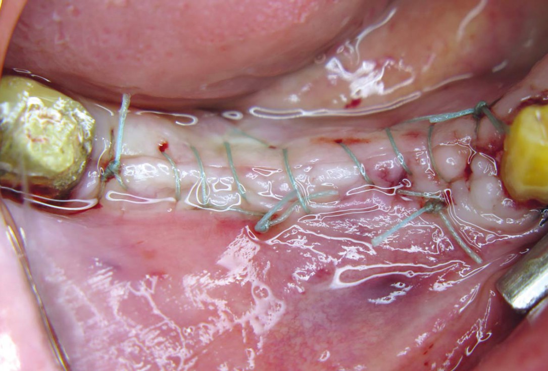
07 / 20 - Tension-free wound closure without compression on the augmentation siteHorizontal ridge augmentation with maxgraft® cortico - M.Sc. E. Kapogianni
-
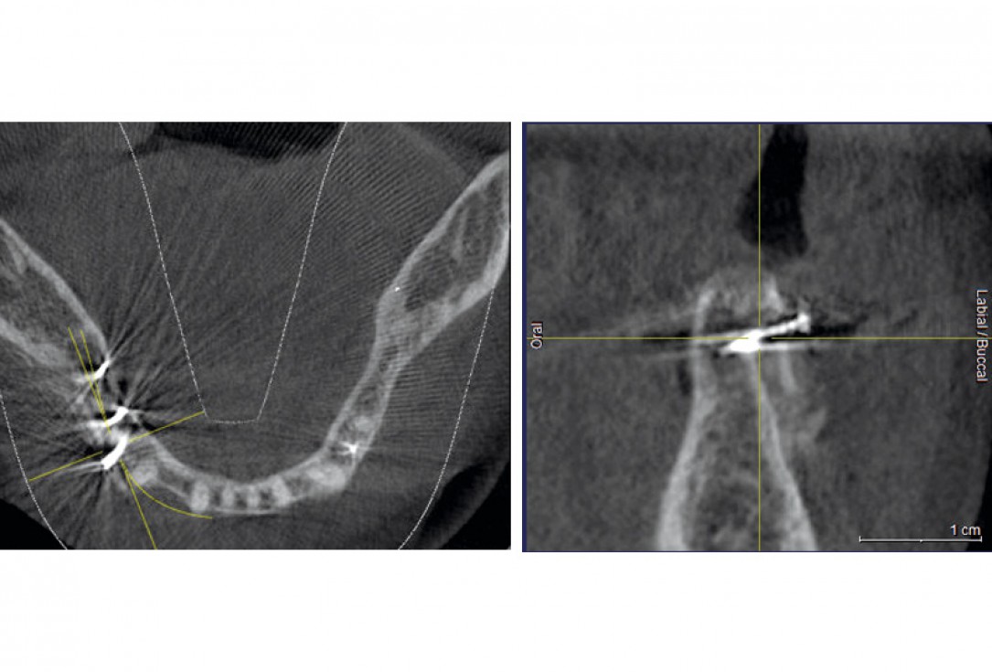
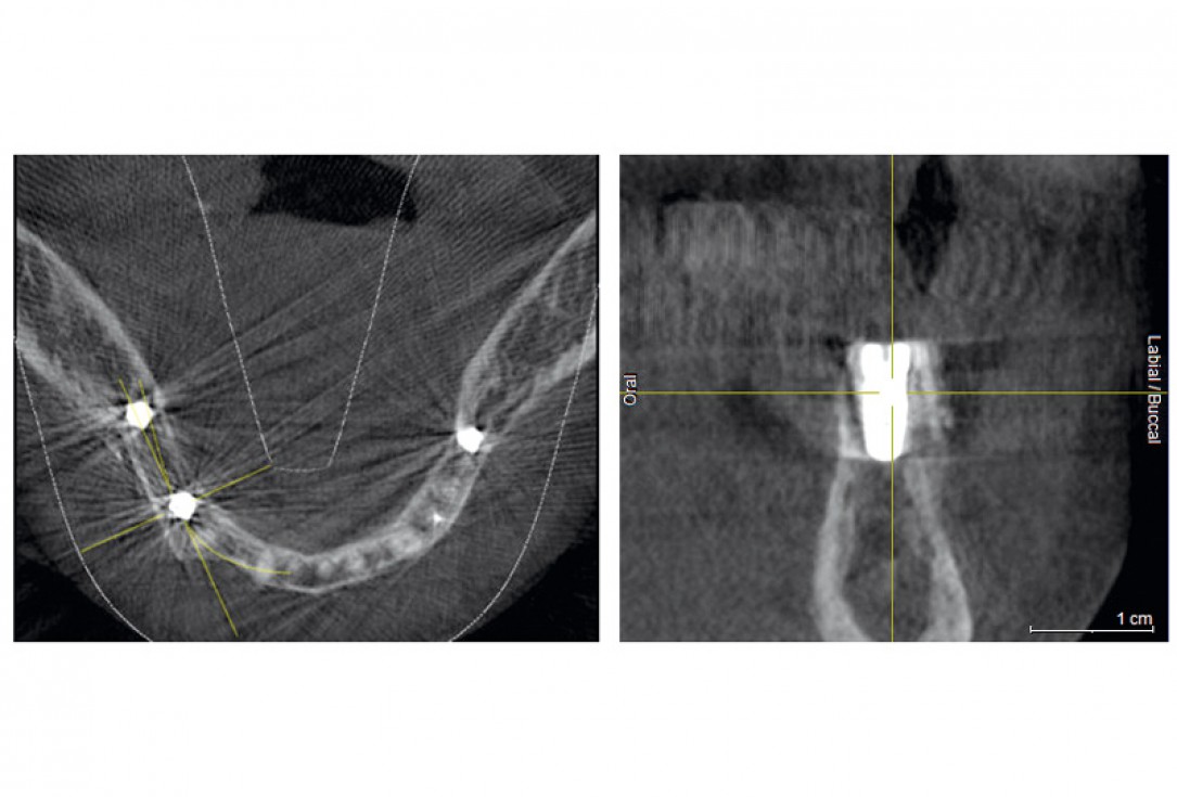
08 / 20 - CBCT recording after the augmentation shows the cortical plate about 3 to 4 mm distant from the host boneHorizontal ridge augmentation with maxgraft® cortico - M.Sc. E. Kapogianni
-
09 / 20 - Reentry after 5 months of healing shows excellent bone regenerationHorizontal ridge augmentation with maxgraft® cortico - M.Sc. E. Kapogianni
-
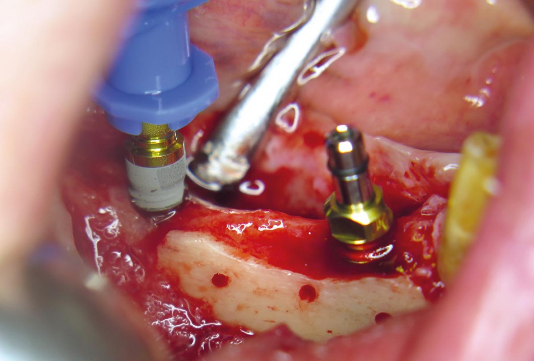
10 / 20 - Placing of pilot drills into the new formed bone tissueHorizontal ridge augmentation with maxgraft® cortico - M.Sc. E. Kapogianni
-
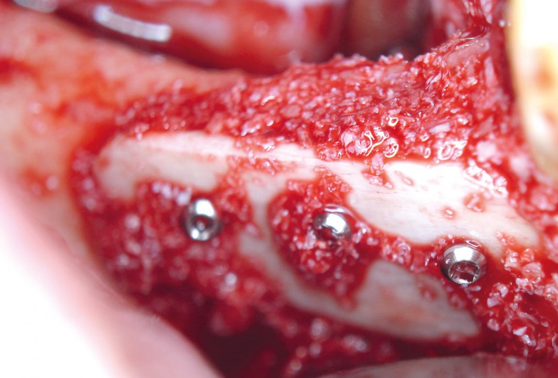
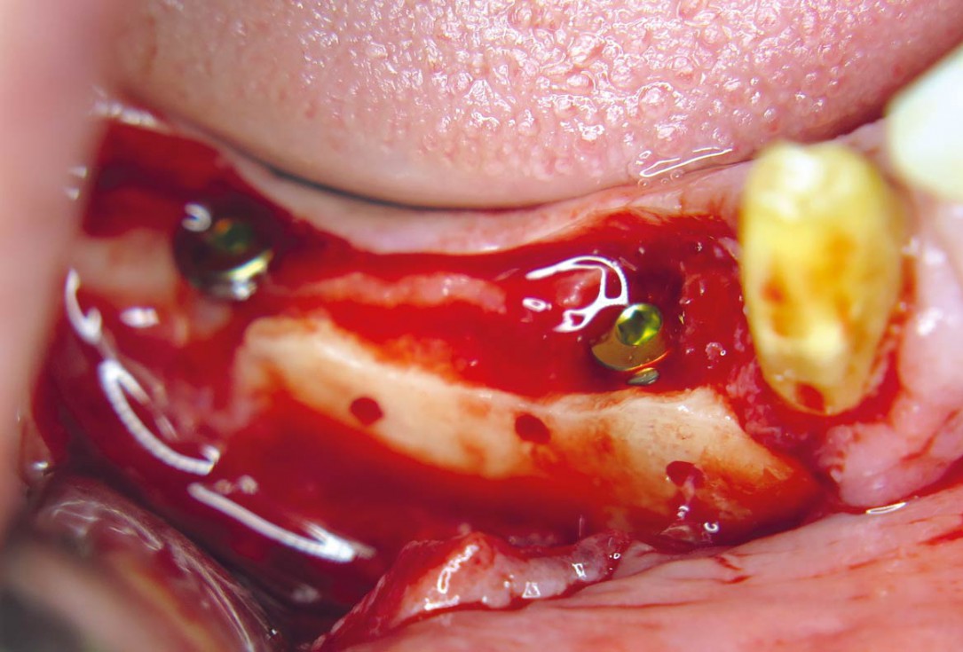
11 / 20 - Insertion of two dental implants in regio 44 and 46Horizontal ridge augmentation with maxgraft® cortico - M.Sc. E. Kapogianni
-
12 / 20 - Stable and fully submerged positioning of implantsHorizontal ridge augmentation with maxgraft® cortico - M.Sc. E. Kapogianni
-
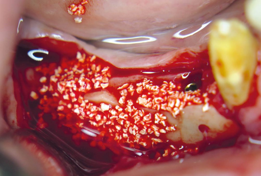
13 / 20 - Contouring with cerabone® for optimal volume stability and an aesthetic outcomeHorizontal ridge augmentation with maxgraft® cortico - M.Sc. E. Kapogianni
-
14 / 20 - Surgical site covering with a collprotect® membraneHorizontal ridge augmentation with maxgraft® cortico - M.Sc. E. Kapogianni
-
15 / 20 - Tension-and compression-free suturesHorizontal ridge augmentation with maxgraft® cortico - M.Sc. E. Kapogianni
-
16 / 20 - CBCT after the implantation shows position of the implant within vital bone tissue and the adjacent allogenic cortical plateHorizontal ridge augmentation with maxgraft® cortico - M.Sc. E. Kapogianni
-
17 / 20 - Soft tissue healing one week after implantationHorizontal ridge augmentation with maxgraft® cortico - M.Sc. E. Kapogianni
-
18 / 20 - Excellent soft tissue situation after three months of healingHorizontal ridge augmentation with maxgraft® cortico - M.Sc. E. Kapogianni
-
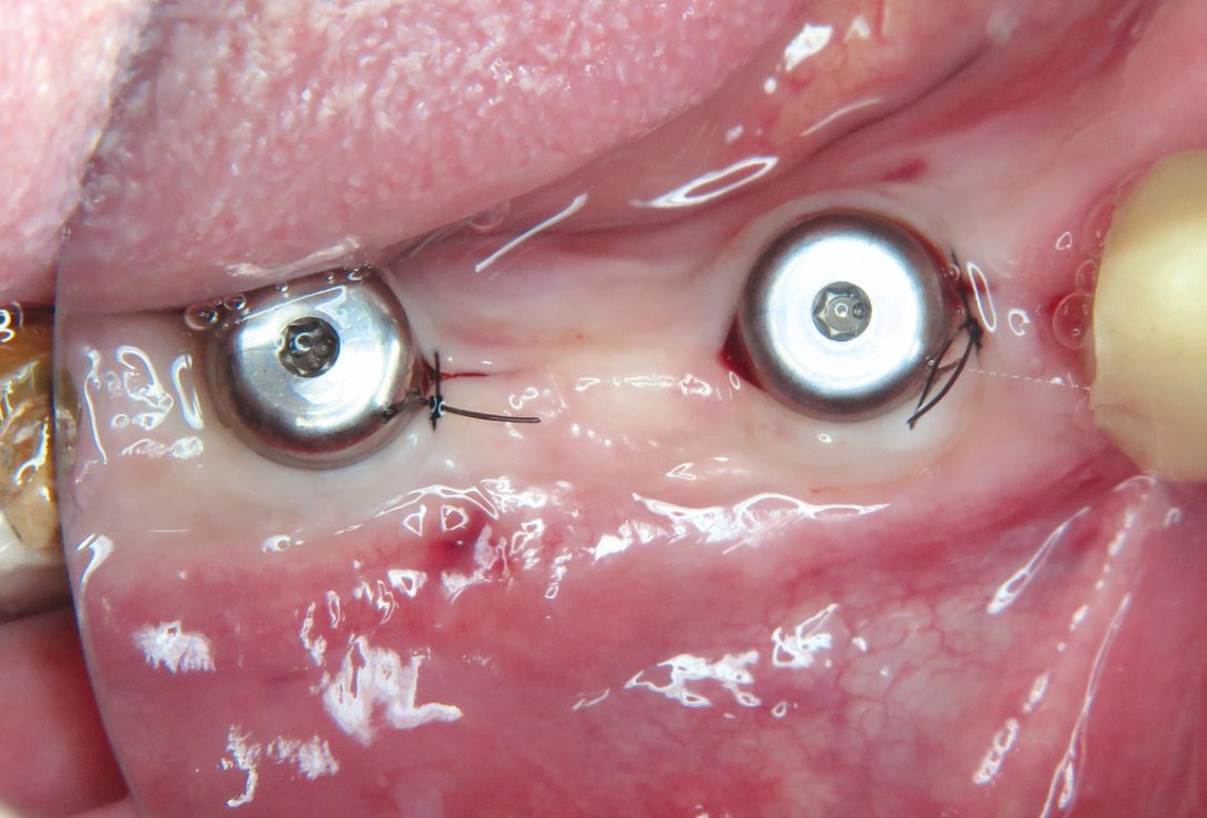
19 / 20 - Placing of gingiva formers for an aesthetic margin between the final crown and the soft tissueHorizontal ridge augmentation with maxgraft® cortico - M.Sc. E. Kapogianni
-
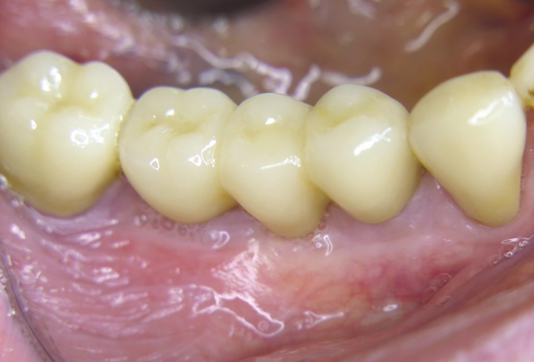
20 / 20 - Final implant-retained denture with natural appearance one month after placing of gingiva formersHorizontal ridge augmentation with maxgraft® cortico - M.Sc. E. Kapogianni

Pre-operative OPG shows deep vertical intrabony defects on the distal aspects of teeth 13 and 14.

Pre-surgical probing reveals a deep intrabony defect on the distal aspect of the upper canine.

Pre-operative radiograph. Intrabony defect on the mesial aspect of tooth 14.

Initial X-ray presenting a very deep intrabony defect of tooth 21

Surgical presentation of the alveolar ridge with reduced amount of horizontal bone available

DVT control after sinusitis surgery, residual bone height 1 mm

OPG of the initial situation – provision of missing denture in regio 44 to 47 by a resin-retained bridge

The patient presented with a terminal fracture of the crown tooth number 12

Clinical situation with narrow alveolar ridge in the lower jaw

Extended horizontal and vertical defect of the maxilla following tumor resection and reconstruction with a scapula graft

Clinical situation of the edentulous distal maxilla before the surgery

DVT image showing the reduced amount of bone available in the area of the mental foramen
