Immediate implant placement in the maxilla with contour GBR - Dr. K. Loukas
-
01/13 - The patient presented with a terminal fracture of the crown tooth number 12Immediate implant placement in the maxilla with contour GBR - Dr. K. Loukas
-
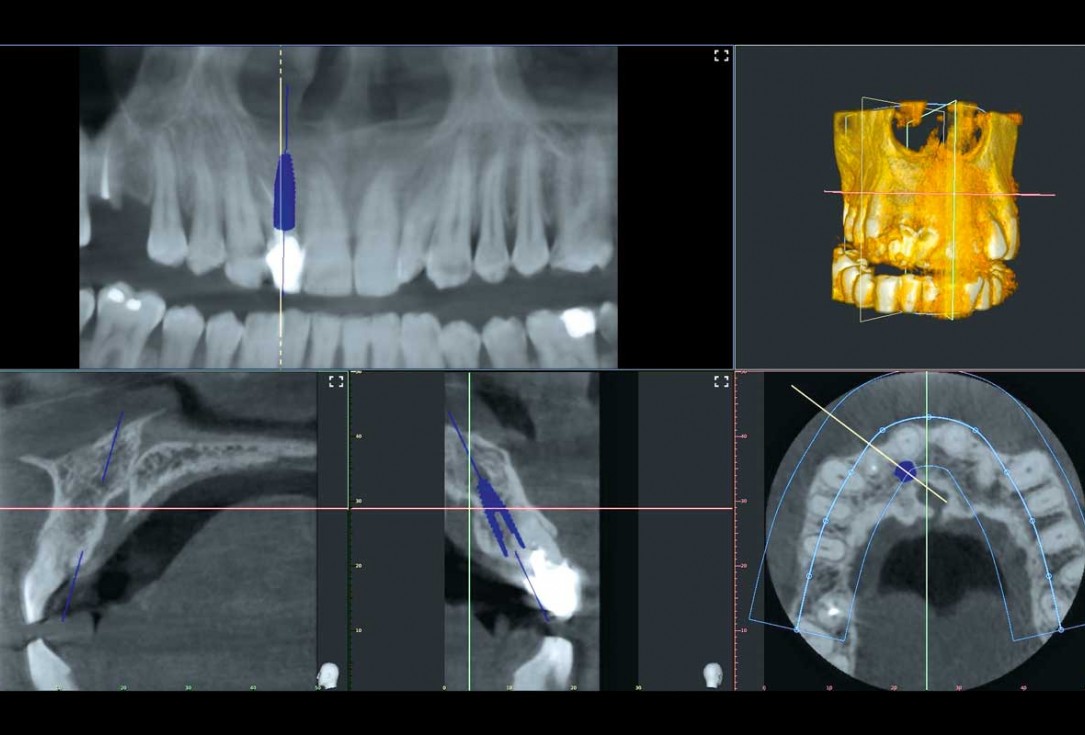
02/13 - The CBCT revealed adequate bone width and no pathology in the area, immediate implant placement was agreedImmediate implant placement in the maxilla with contour GBR - Dr. K. Loukas
-
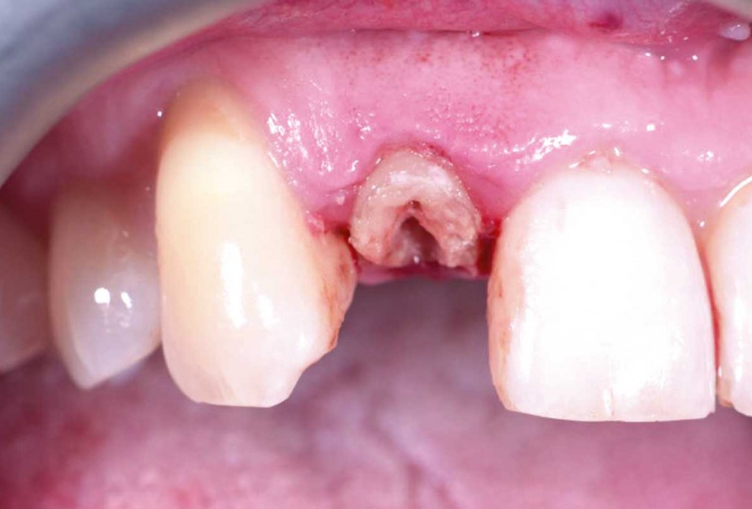
03/13 - Root condition, gingival zenith higher than the 11, intact distal papilla and some recession in mesial papillaImmediate implant placement in the maxilla with contour GBR - Dr. K. Loukas
-
04/13 - Vertical extraction of tooth 12Immediate implant placement in the maxilla with contour GBR - Dr. K. Loukas
-
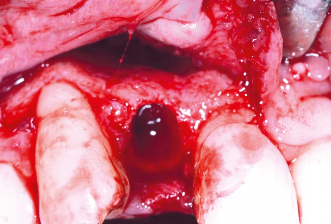
05/13 - Full muco-periostal flap elevationImmediate implant placement in the maxilla with contour GBR - Dr. K. Loukas
-
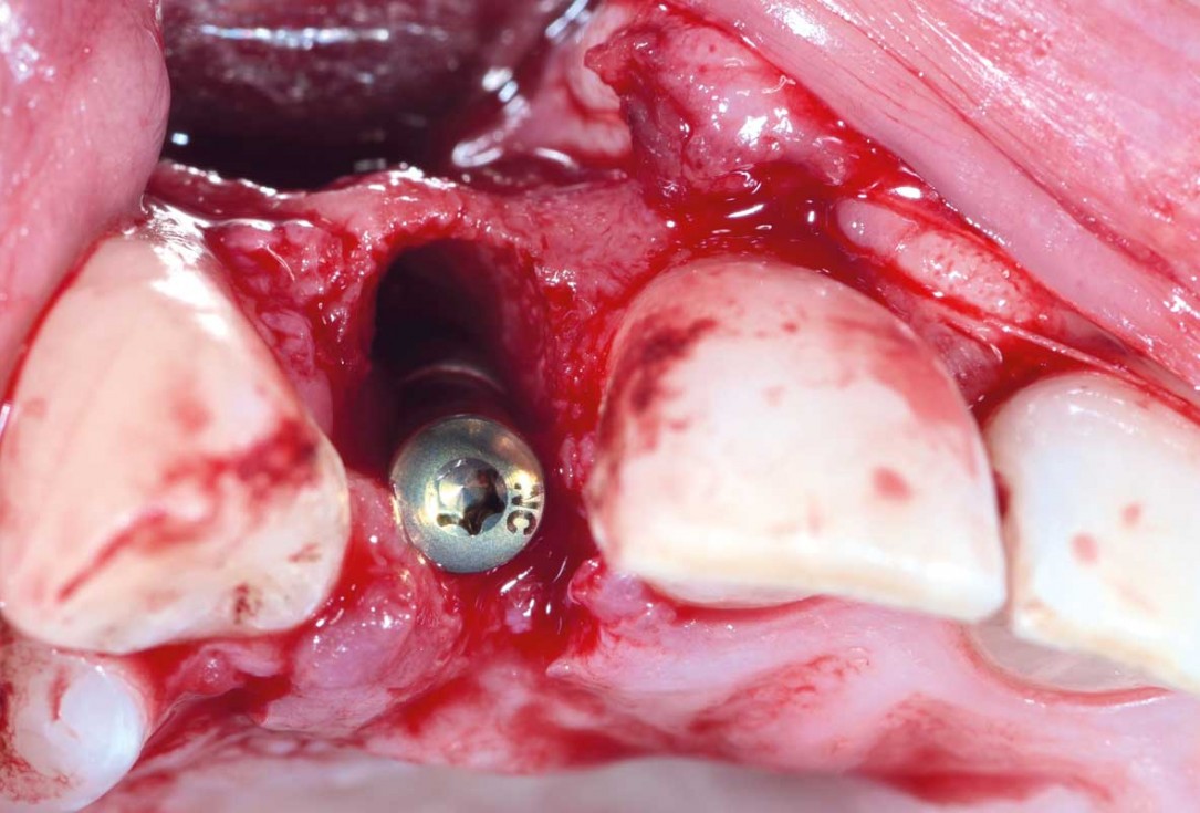
06/13 - Implant (3.3 BL x 12 mm) inserted in correct 3D prosthetic position, a 3 mm distance gap was left in the crestal section; Compared to CBCT apical concavity was more intenseImmediate implant placement in the maxilla with contour GBR - Dr. K. Loukas
-
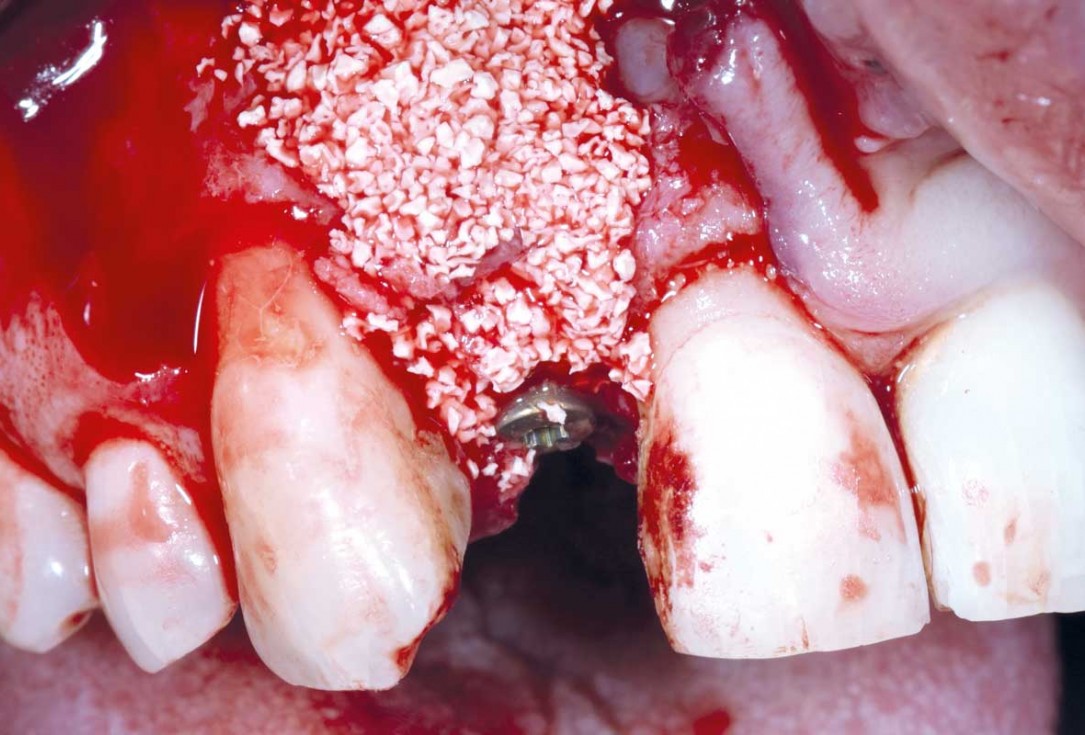
07/13 - Autogenous chips applied in contact with the implant, cerabone® mixed with autogenous chips was applied inside and outside; A long conical healing abutment was used in situImmediate implant placement in the maxilla with contour GBR - Dr. K. Loukas
-
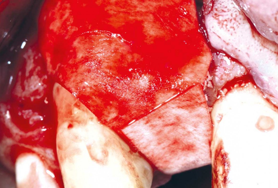
08/13 - Application of a double layer of the resorbable collprotect® membraneImmediate implant placement in the maxilla with contour GBR - Dr. K. Loukas
-
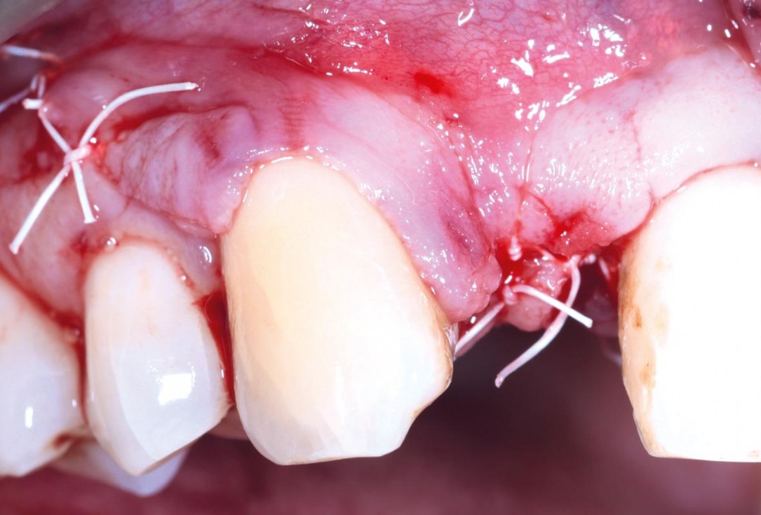
09/13 - Closure with PTFE suturesImmediate implant placement in the maxilla with contour GBR - Dr. K. Loukas
-
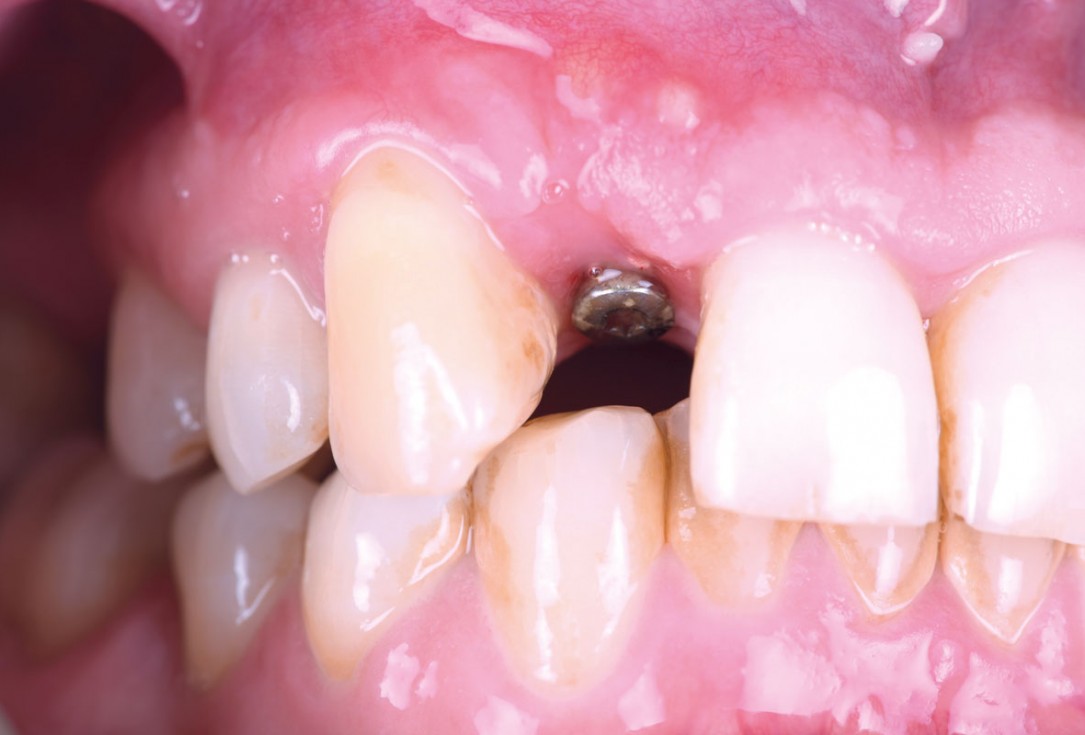
10/13 - Clinical view after 8 weeks of healingImmediate implant placement in the maxilla with contour GBR - Dr. K. Loukas
-
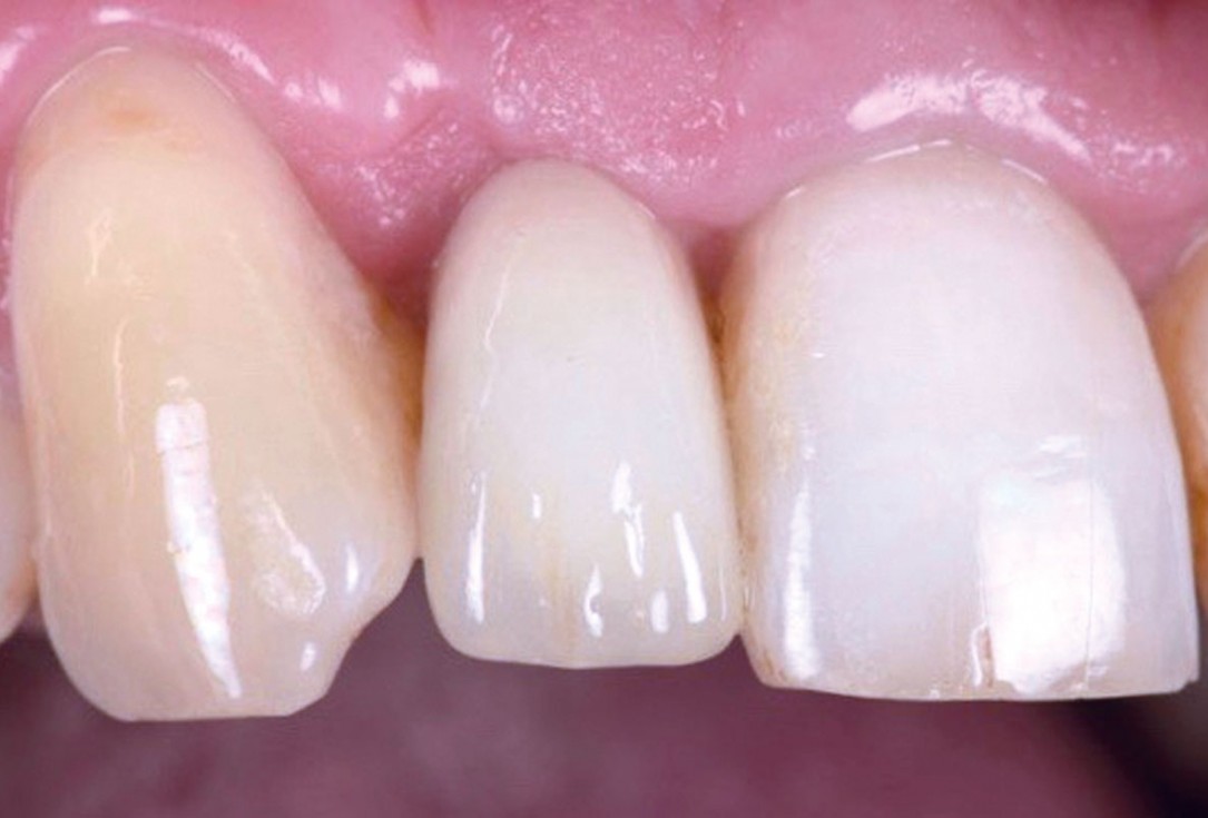
11/13 - After 10 weeks delivery of the screw retained crown with a CAD CAM milled abutmentImmediate implant placement in the maxilla with contour GBR - Dr. K. Loukas
-
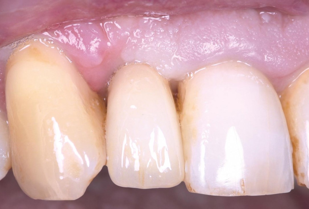
12/13 - Clinical situation 4 months post surgery: Excellent hard and soft tissue adaptation, papilla closing bilaterallyImmediate implant placement in the maxilla with contour GBR - Dr. K. Loukas
-
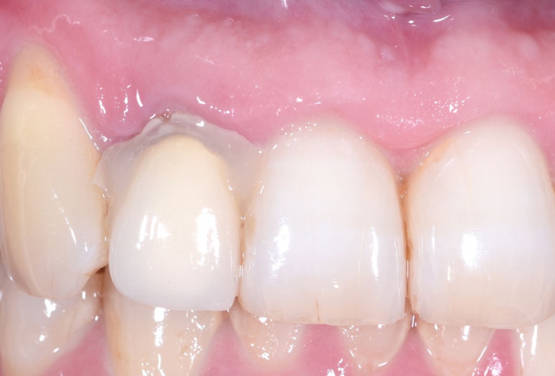
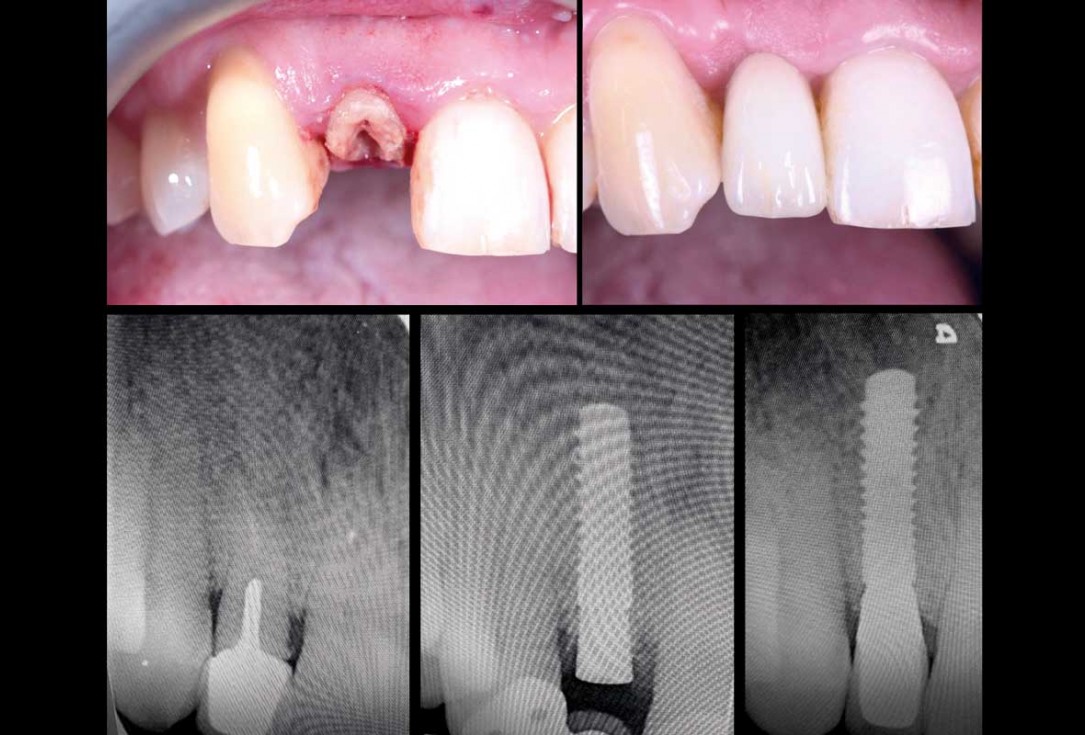
13/13 - Before and after comparison: Clinical and radiologic viewImmediate implant placement in the maxilla with contour GBR - Dr. K. Loukas

Initial clinical situation. Atrophic maxillary ridge.

Initial x-ray showing bone loss around implants placed 5 years ago in another dental clinic

Initial view of the case. Discoloration of 1.1 and mild class I gingival recession

Situation after tooth removal.

Initial clinical situation with gum recession and labial bone loss eight weeks following tooth extraction

Three implants placed in a narrow posterior mandible

Pre-operative clinical situation.

Clinical situation with narrow alveolar ridge in the lower jaw

Initial clinical situation showing bone wall defect.

Initial clinical situation.

Pre-surgical situation.

Initial situation: missing teeth #11 & 12 and badly broken #21 root

Pre-operative OPG shows deep vertical intrabony defects on the distal aspects of teeth 13 and 14.

Instable bridge situation with abscess formation at tooth #15 after apicoectomy

Initial clinical situation.

Implant insertion in atrophic alveolar ridge

Preoperative clinical situation

Pre-operative OPG

Pre-operative X-ray. Hopless tooth 21.

Pre-surgical situation. Teeth 26 and 27 missing.

Extraction of tooth 21 after endodontic treatment

Pre-surgical probing reveals a deep intrabony defect on the distal aspect of the upper canine.

Initial clinical situation with single tooth gap in regio 21

Pre-operative radiographic view. Intrabony defect on the distal aspect of the lateral incisor.

Clinical situation before extraction and implantation

Pre-operative radiographic view.
