Isolated gingival recession treated with the modified coronally advanced tunnel in conjunction with Straumann® Emdogain® and autologous CTG - Dr. R. Cosgarea
-
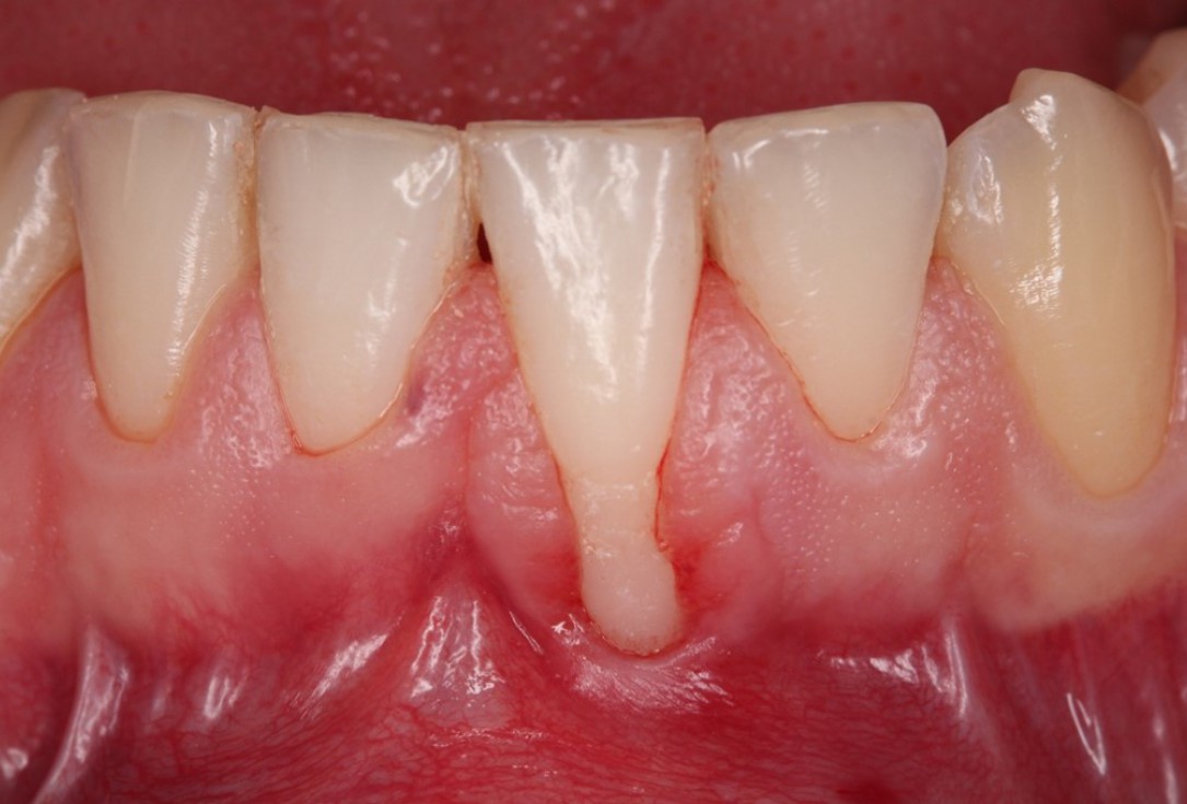
01/06 - Baseline clinical situation. Recession depth of 6 mm at tooth 31.Isolated gingival recession treated with the modified coronally advanced tunnel in conjunction with Straumann® Emdogain® and autologous CTG - Dr. R. Cosgarea
-
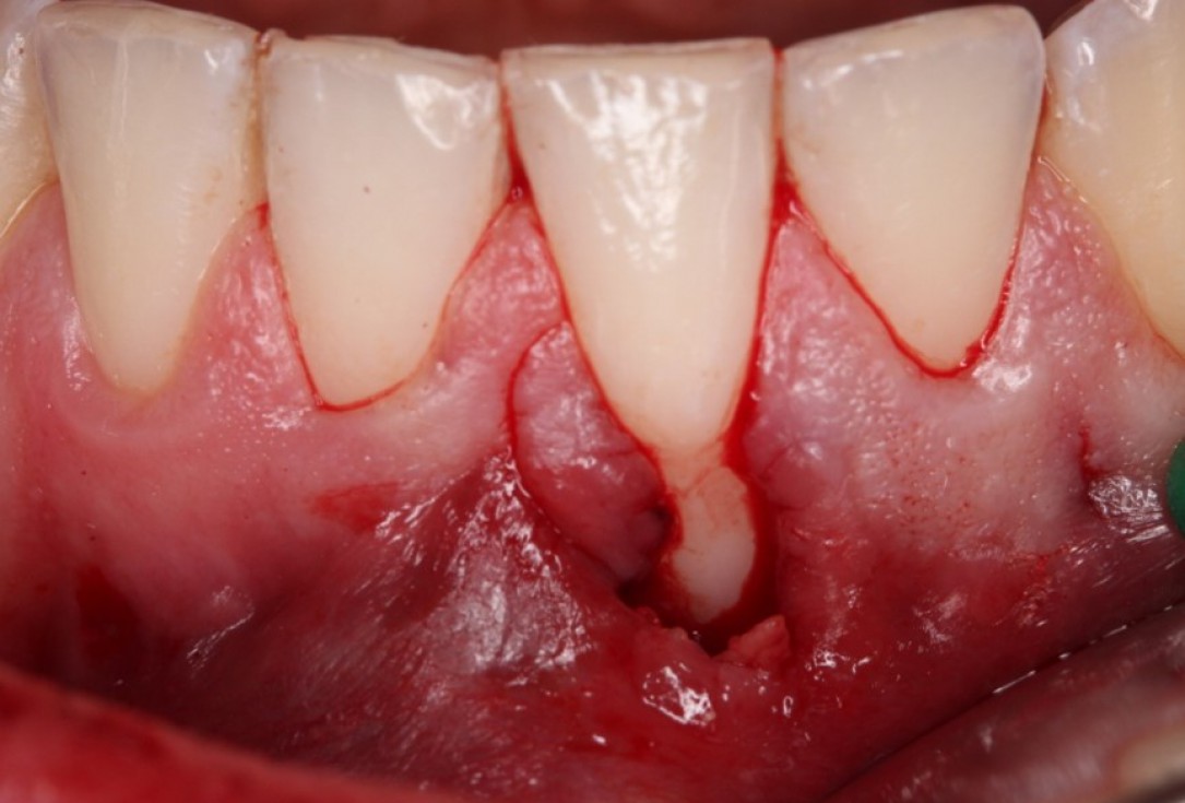
02/06 - Preparation of the tunnel.Isolated gingival recession treated with the modified coronally advanced tunnel in conjunction with Straumann® Emdogain® and autologous CTG - Dr. R. Cosgarea
-
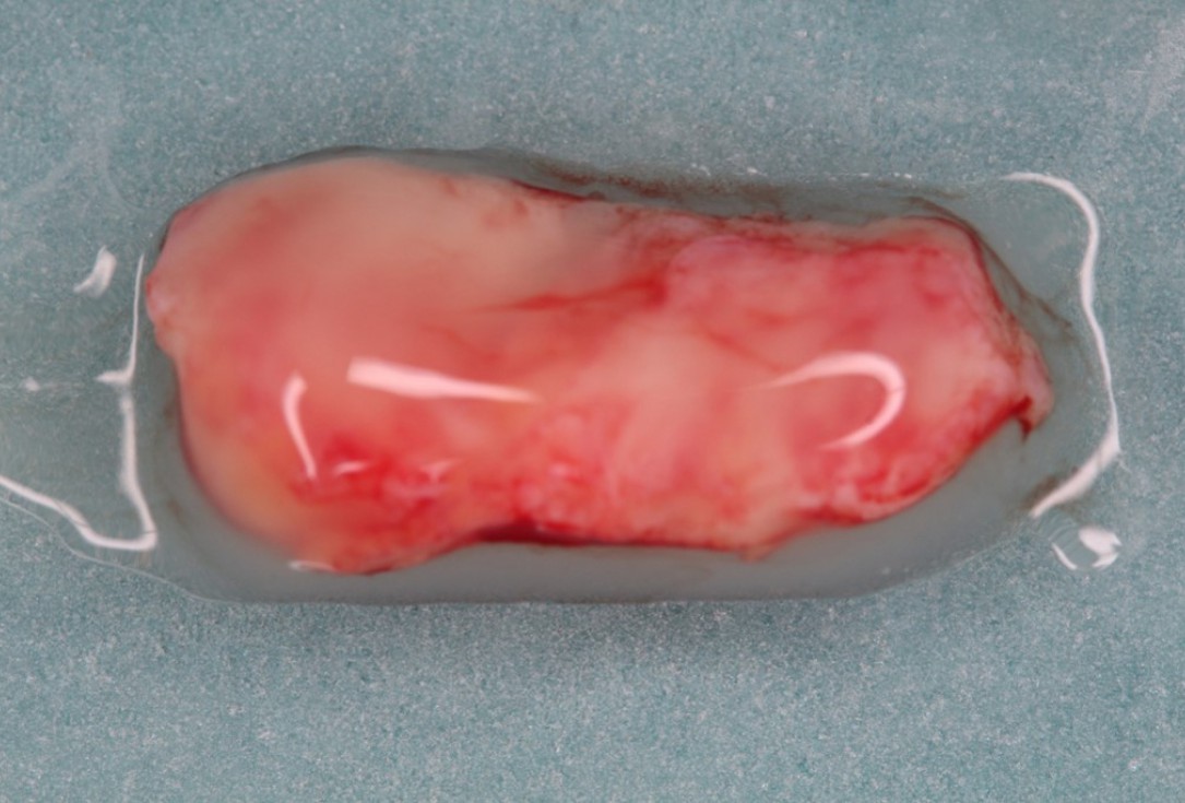
03/06 - Connective tissue graft (CTG) coated with Straumann® Emdogain®.Isolated gingival recession treated with the modified coronally advanced tunnel in conjunction with Straumann® Emdogain® and autologous CTG - Dr. R. Cosgarea
-
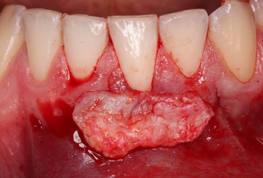
04/06 - Adaptation of the CTG.Isolated gingival recession treated with the modified coronally advanced tunnel in conjunction with Straumann® Emdogain® and autologous CTG - Dr. R. Cosgarea
-
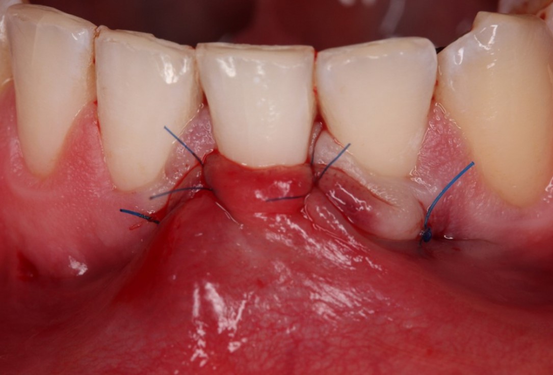
05/06 - Suturing.Isolated gingival recession treated with the modified coronally advanced tunnel in conjunction with Straumann® Emdogain® and autologous CTG - Dr. R. Cosgarea
-
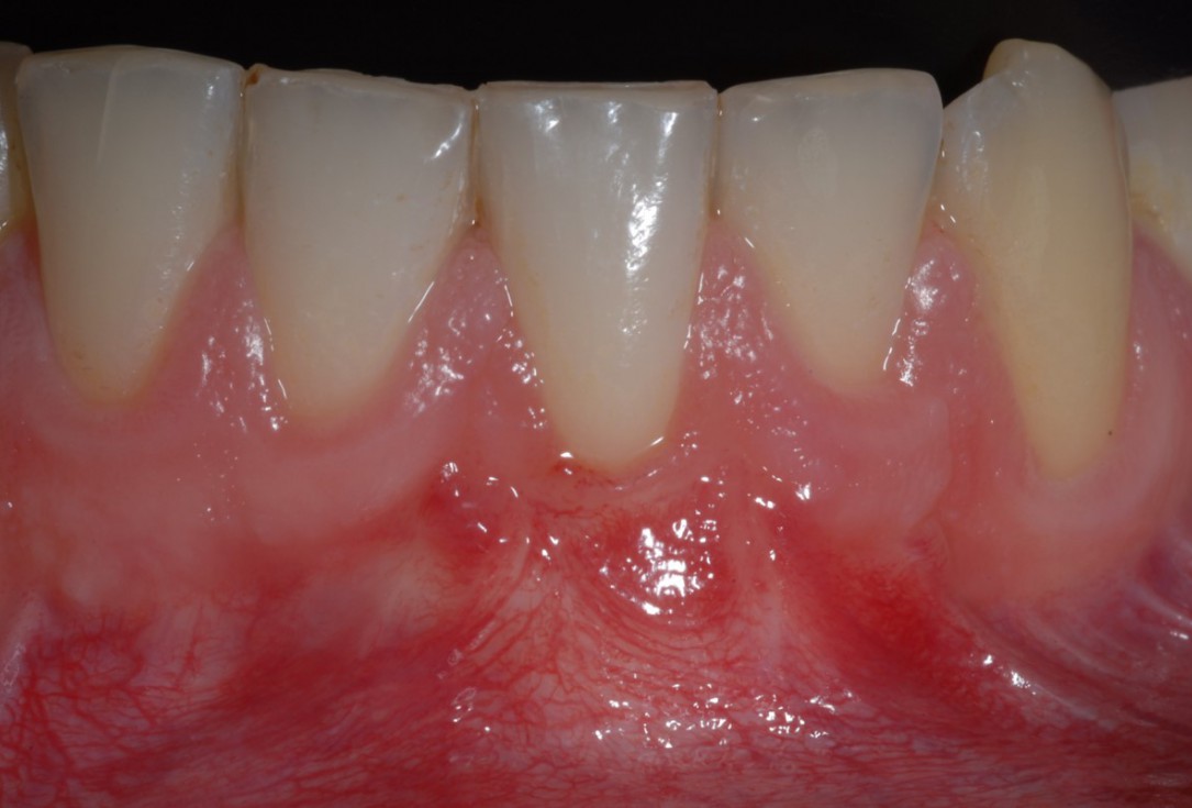
06/06 - Clinical situation 1 year post-operative.Isolated gingival recession treated with the modified coronally advanced tunnel in conjunction with Straumann® Emdogain® and autologous CTG - Dr. R. Cosgarea

Alveolar socket before soft and hard tissue augmentation

Pre-operative OPG shows deep vertical intrabony defects on the distal aspects of teeth 13 and 14.

Radiographic view before periodontal regenerative therapy with Straumann® Emdogain®. A deep intrabony defect appeared mesially and distally on the left mandibular first premolar. Pre-surgical probing measured 8 mm. The defect morphology presented as well-contained.

Baseline clincial situation and pre-surgical probing.

Pre-operative radiographic view.

Pre-surgical situation. Multiple adjacent gingival recessions at teeth 12, 13 and 14.

Pre-operative clinical view. Multiple adjacent gingival recessions.

Initial clinical situation

Pre-operative X-ray. Hopless tooth 21.

Pre-operative clinical situation.

Pre-operative radiograph. Intrabony defect on the mesial aspect of tooth 14.

Pre-operative clinical situation. Gingival recessions at teeth 11 and 21.

Pre-operative clinical situation. Multiple adjacent gingival recessions.

Pre-operative clinical situation. Shallow multiple adjacent gingival recessions in the first quadrant.

Initial situation: bone loss due to lack of physical load of bridge retained region 11

Pre-operative radiographic view. Intrabony defect on the distal aspect of the lateral incisor.

Situation after tooth removal.

Pre-operative clinical situation.

Pre-surgical clinical situation, buccal view.

Pre-operative probing pocket depth (PPD) at the distal aspect of tooth 11 was 7 mm.

Pre-surgical clinical situation. Deep gingival recessions at both upper canine.

Initial situation: 40 year old female patient with extensive scar tissue after several surgeries restored with a Rochette bridge
