Three-dimensional augmentation with maxgraft® cortico - Dr. R. Würdinger
-
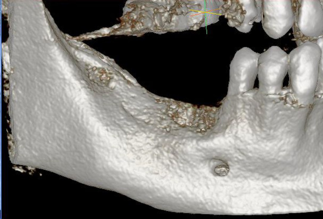
01/28 - Model of the initial defect computed from a CBCT scan - buccal viewThree-dimensional augmentation with maxgraft® cortico - Dr. R. Würdinger
-
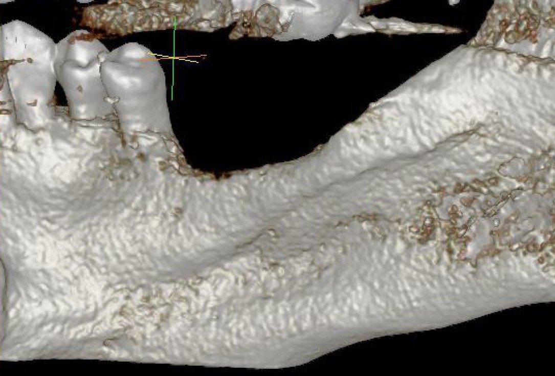
02/28 - Model of the initial defect computed from a CBCT scan - lingual viewThree-dimensional augmentation with maxgraft® cortico - Dr. R. Würdinger
-
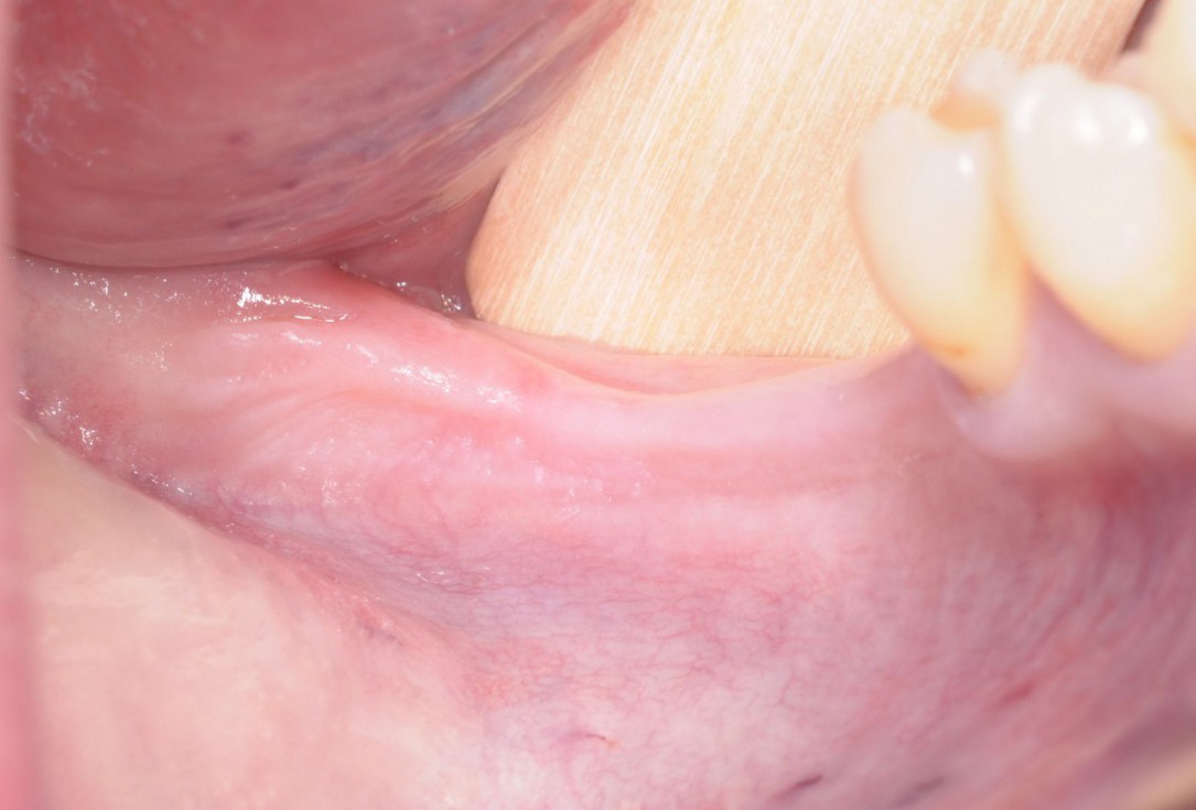
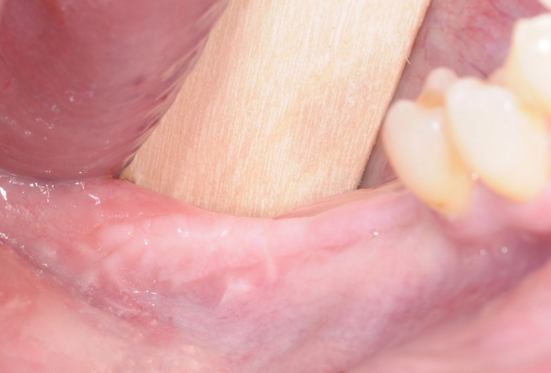
03/28 - Initial clinical situation - massive bone loss in the posterior 4th quadrantThree-dimensional augmentation with maxgraft® cortico - Dr. R. Würdinger
-
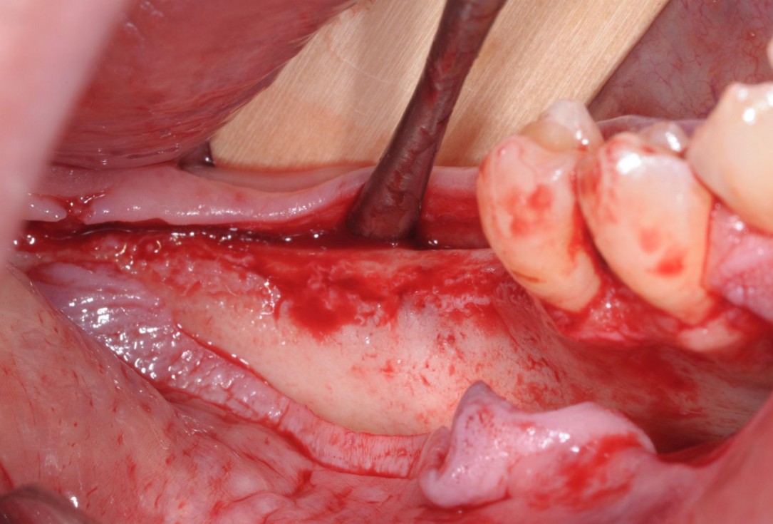
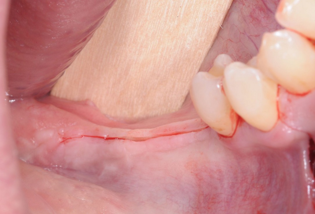
04/28 - Full-thickness flap preparation via crestal incisionThree-dimensional augmentation with maxgraft® cortico - Dr. R. Würdinger
-
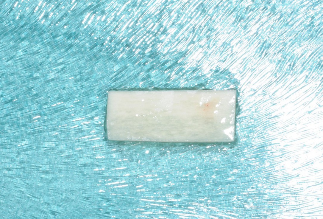
05/28 - maxgraft® cortico rehydrated in saline solution for 30 minutesThree-dimensional augmentation with maxgraft® cortico - Dr. R. Würdinger
-
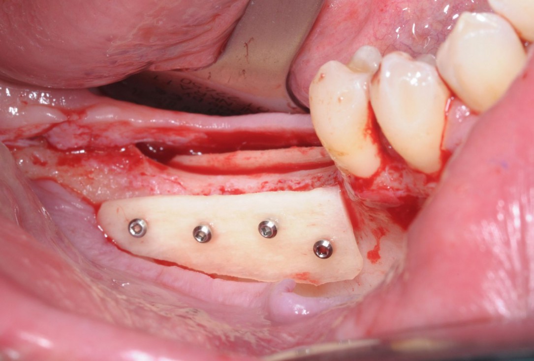
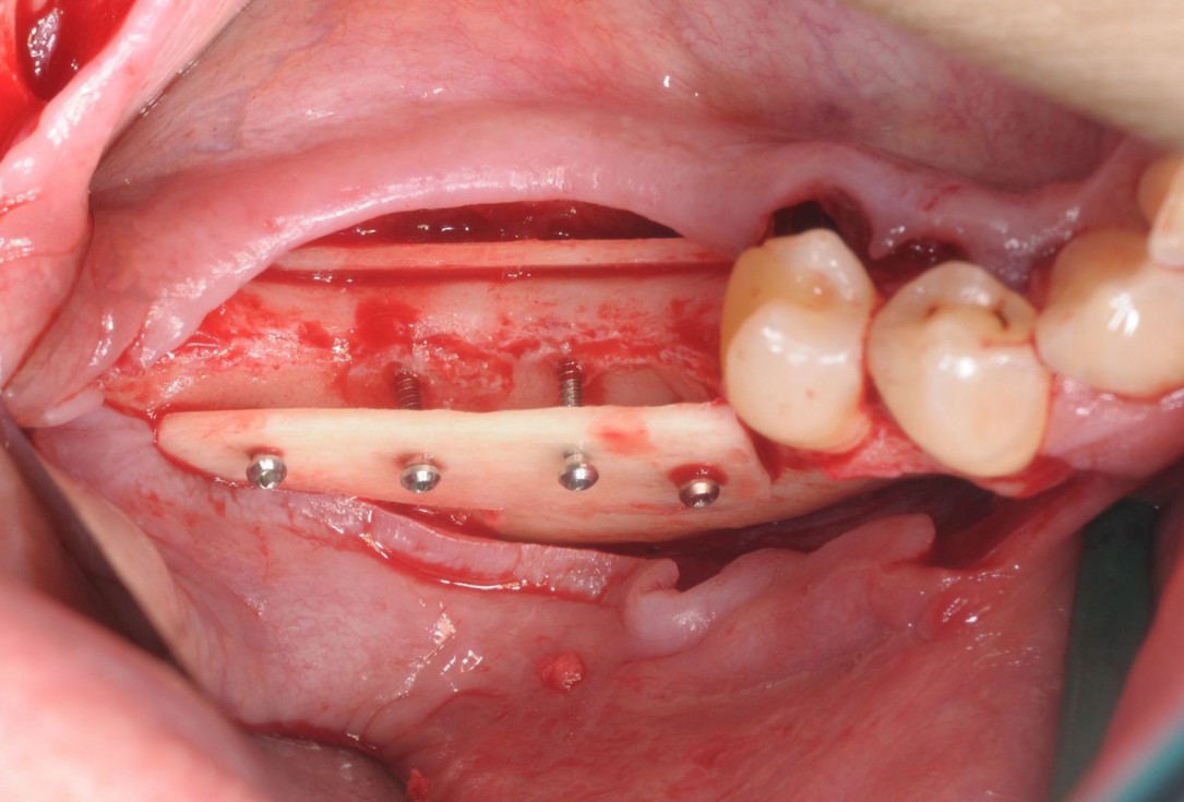
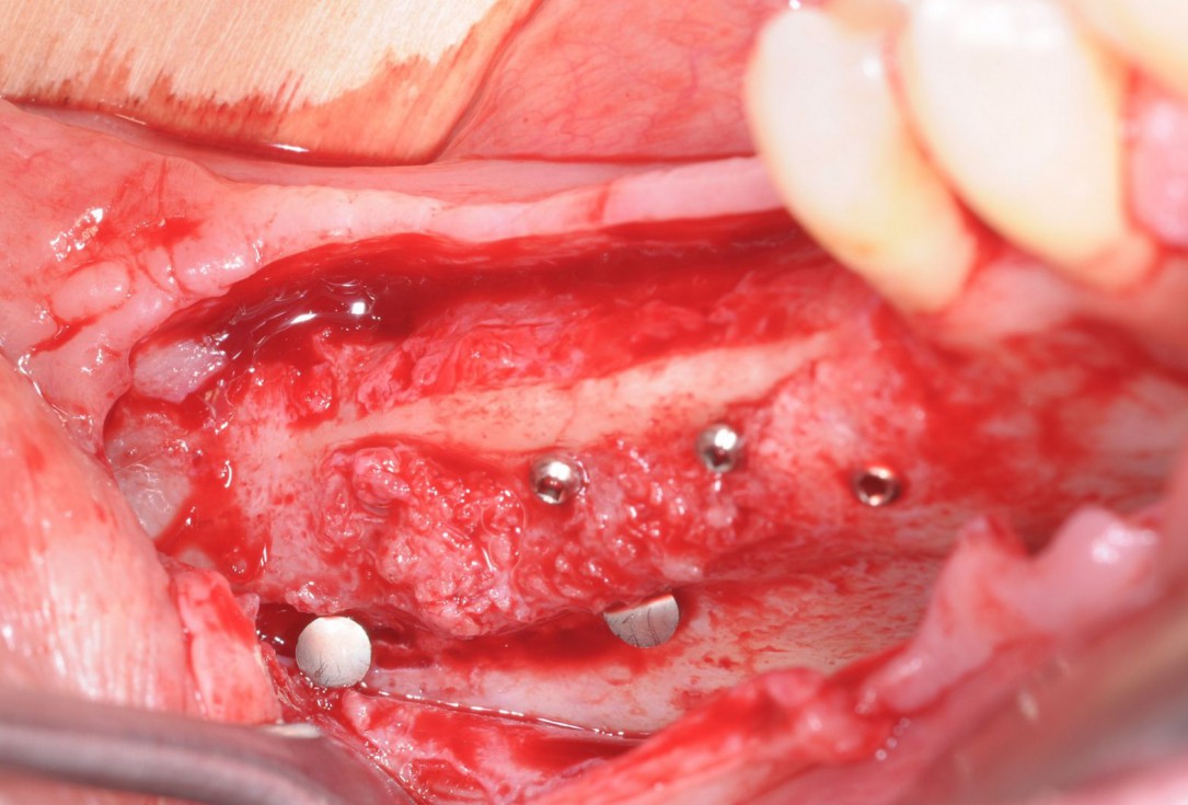
06/28 - Fixation of two defect-adapted maxgraft® cortico plates with osteosynthesis screwsThree-dimensional augmentation with maxgraft® cortico - Dr. R. Würdinger
-
07/28 - Occlusal view on the immobile container created with the two cortical platesThree-dimensional augmentation with maxgraft® cortico - Dr. R. Würdinger
-
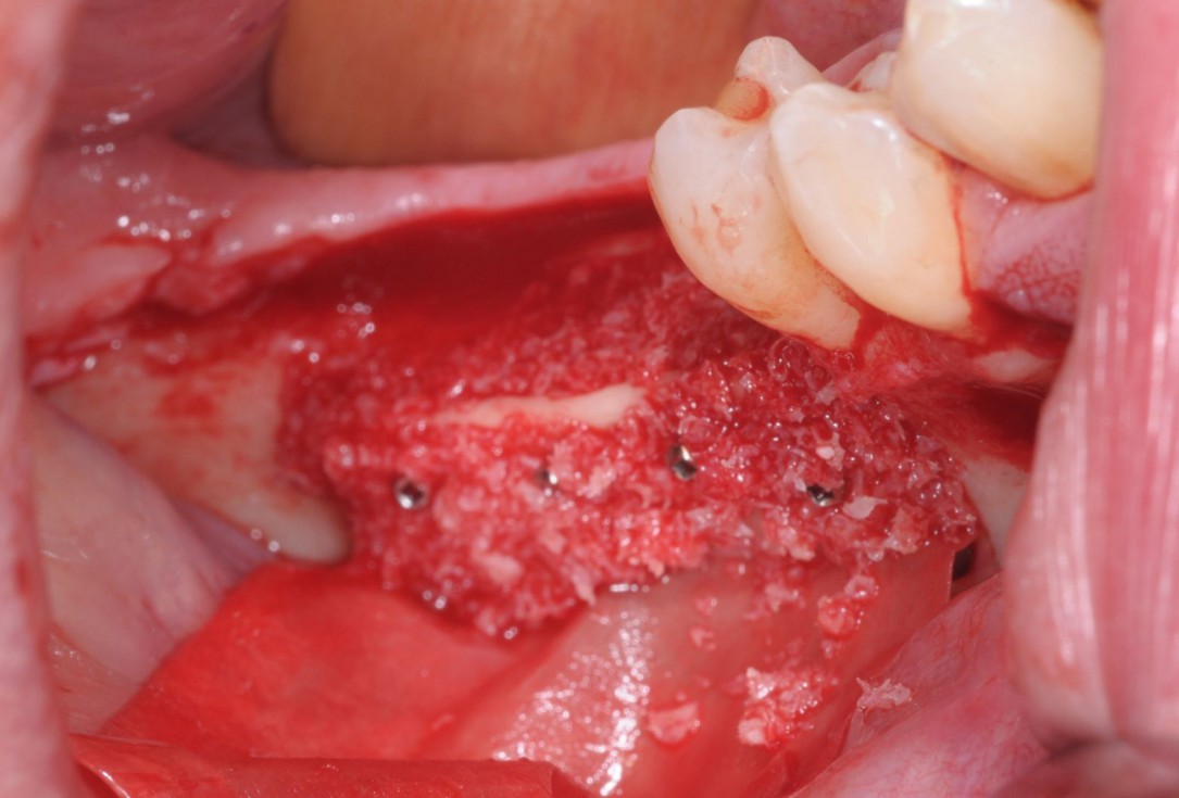
08/28 - Defect-fill and contouring with a composite of autologous and allogenic maxgraft® granules and covering with Jason® membraneThree-dimensional augmentation with maxgraft® cortico - Dr. R. Würdinger
-
09/28 - Application of L-PRF matrices for improved wound healingThree-dimensional augmentation with maxgraft® cortico - Dr. R. Würdinger
-
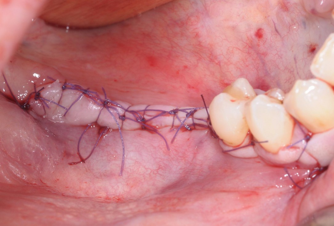
10/28 - Saliva-tight and compression-free wound closure with single-button and mattress suturesThree-dimensional augmentation with maxgraft® cortico - Dr. R. Würdinger
-
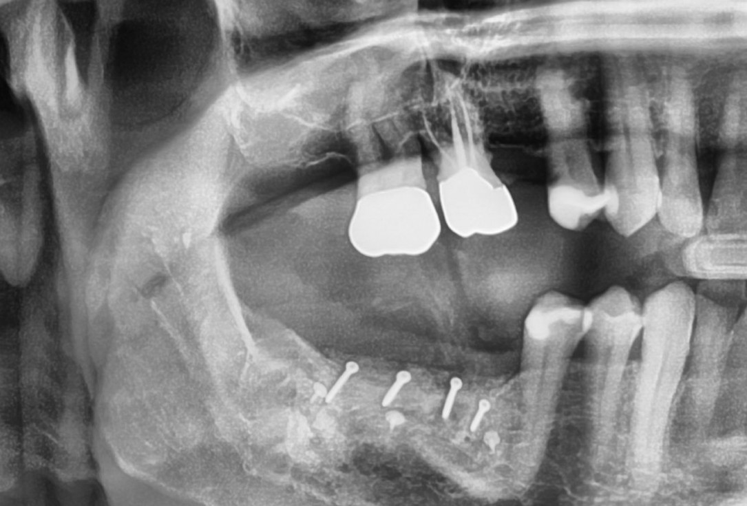
11/28 - X-ray scan after augmentationThree-dimensional augmentation with maxgraft® cortico - Dr. R. Würdinger
-
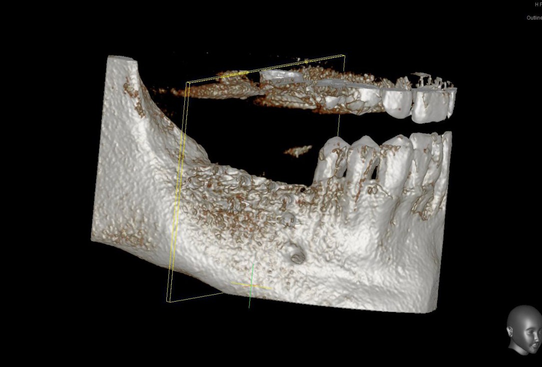
12/28 - Model of the augmented ridge before implantation computed from CBCT-recordings - buccal viewThree-dimensional augmentation with maxgraft® cortico - Dr. R. Würdinger
-
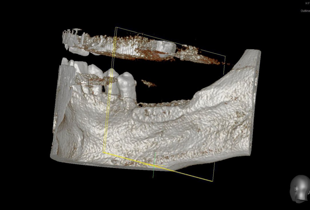
13/28 - Model of the augmented ridge before implantation computed from CBCT-recordings - lingual viewThree-dimensional augmentation with maxgraft® cortico - Dr. R. Würdinger
-
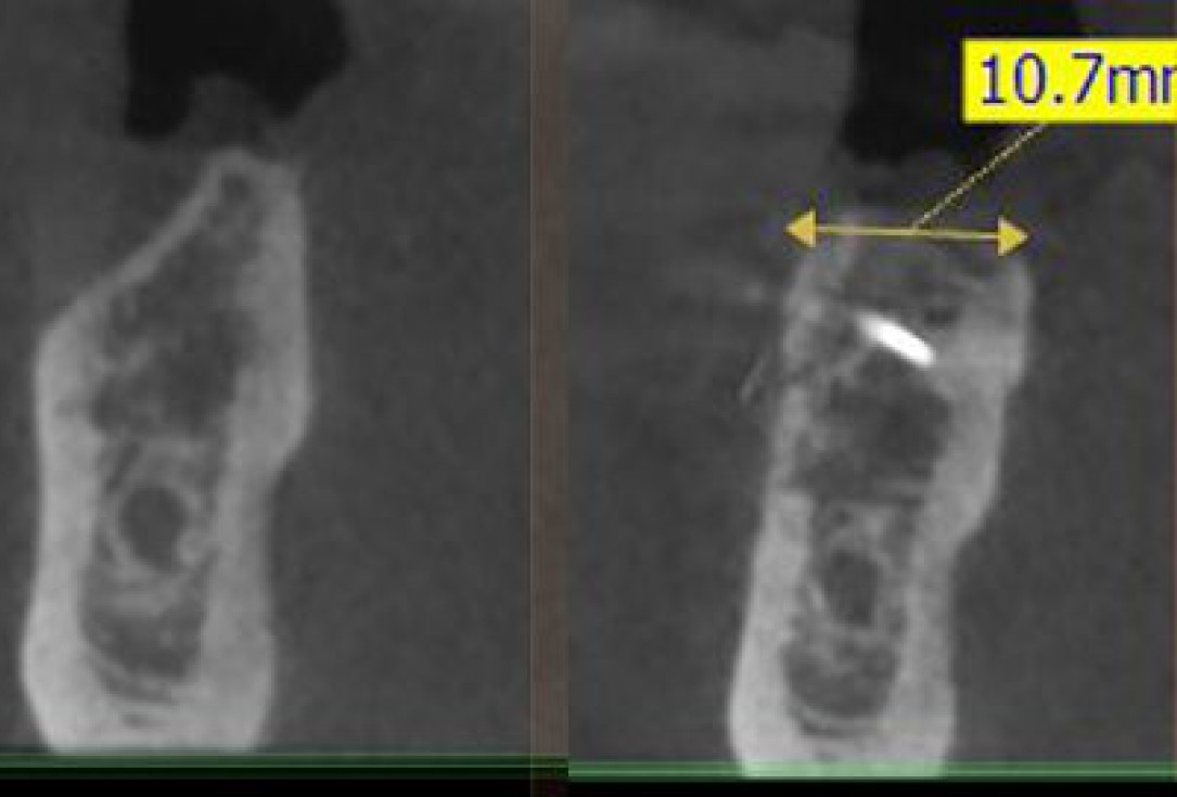
14/28 - Sagittal section of the alveolar ridge in the region of the defect before the augmentation and after 5 months of healing at implantationThree-dimensional augmentation with maxgraft® cortico - Dr. R. Würdinger
-
15/28 - Clinical situation 5 months after ridge augmentationThree-dimensional augmentation with maxgraft® cortico - Dr. R. Würdinger
-
16/28 - Surgical site opening along the initial incision lines to prevent further scar tissue formationThree-dimensional augmentation with maxgraft® cortico - Dr. R. Würdinger
-
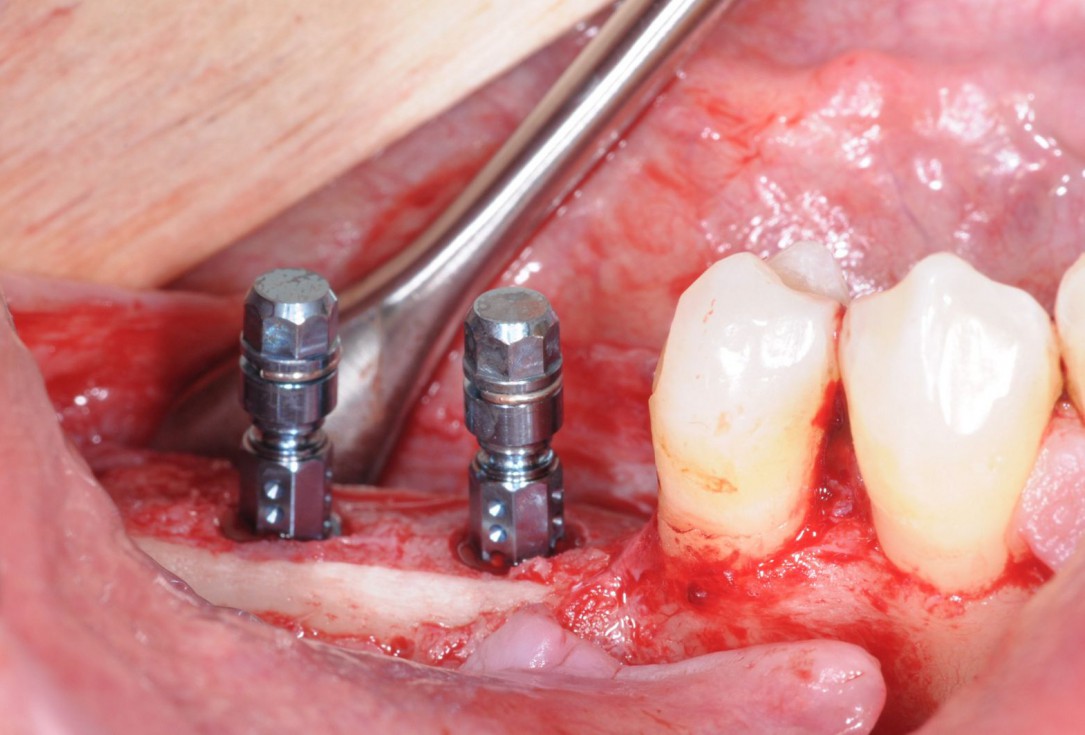
17/28 - Excellent osseous integration of the allogenic cortical plates with new bone attached to the outside and optimal bone tissue regenerationThree-dimensional augmentation with maxgraft® cortico - Dr. R. Würdinger
-
18/28 - Implantation of two Straumann BLT implants into the augmented boneThree-dimensional augmentation with maxgraft® cortico - Dr. R. Würdinger
-
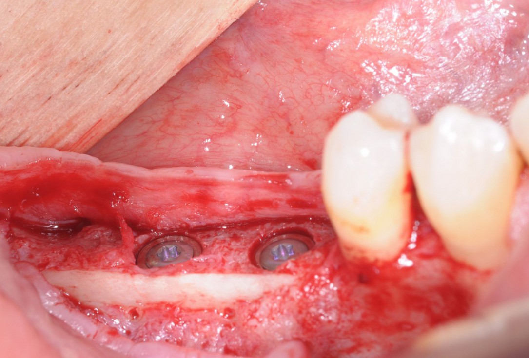
19/28 - Placement of implant cover screws in the fully submerged dental implantsThree-dimensional augmentation with maxgraft® cortico - Dr. R. Würdinger
-
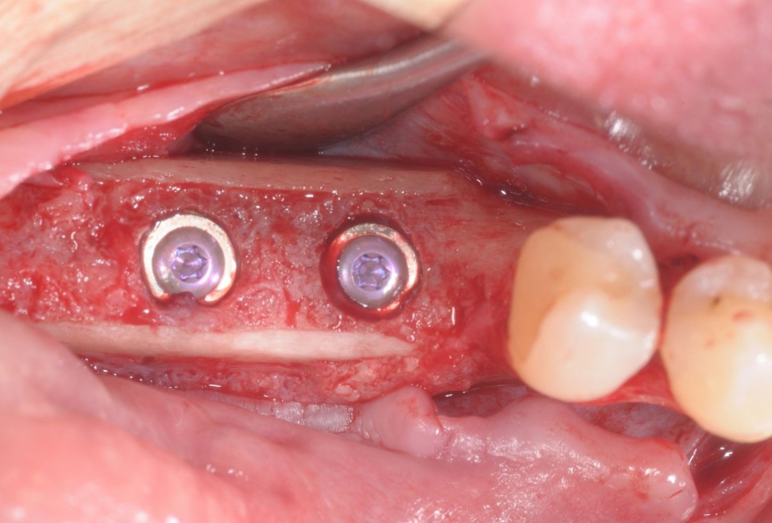
20/28 - Occlusal view of the implants with cover screwsThree-dimensional augmentation with maxgraft® cortico - Dr. R. Würdinger
-
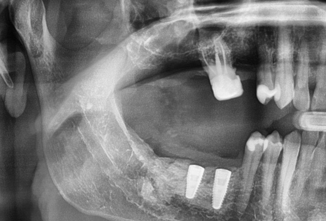
21/28 - X-ray scan of the augmented ridge after implantationThree-dimensional augmentation with maxgraft® cortico - Dr. R. Würdinger
-
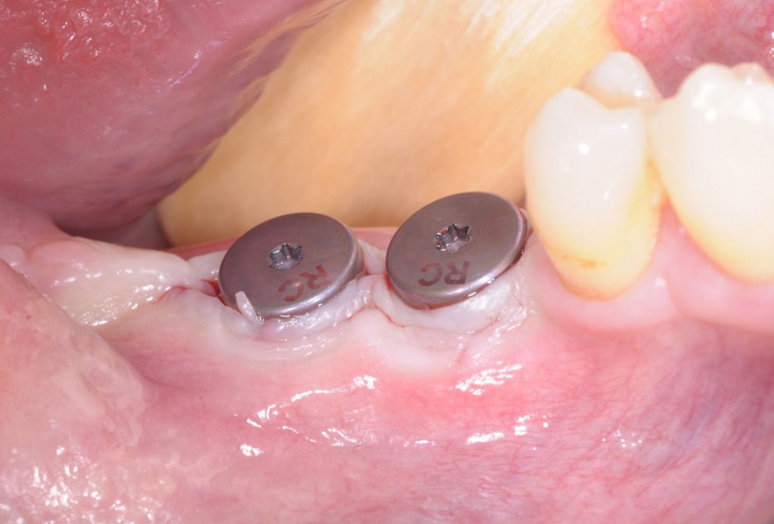
22/28 - Stab incision for reopening of the surgical site and installation of gingival formers - buccal viewThree-dimensional augmentation with maxgraft® cortico - Dr. R. Würdinger
-
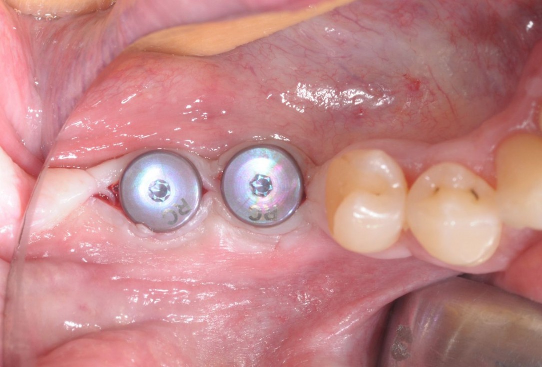
23/28 - Stab incision for reopening of the surgical site and installation of gingival formers - occlusal viewThree-dimensional augmentation with maxgraft® cortico - Dr. R. Würdinger
-
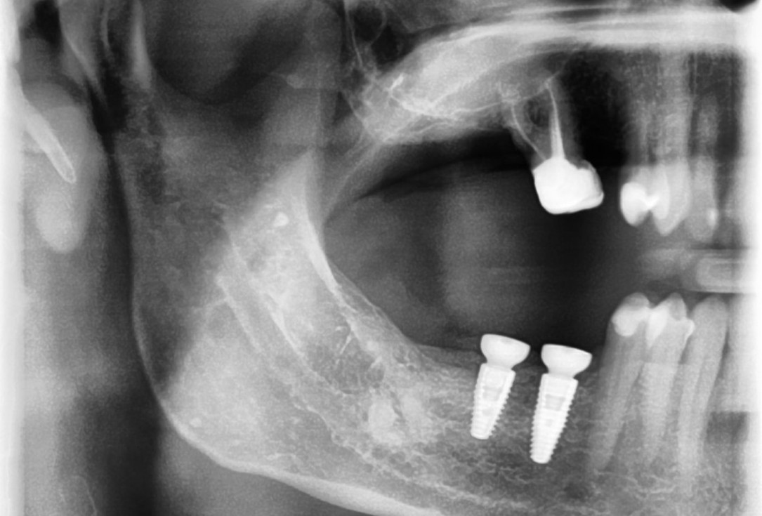
24/28 - X-ray control scan after implant uncovering - excellent and stable osseous situationThree-dimensional augmentation with maxgraft® cortico - Dr. R. Würdinger
-
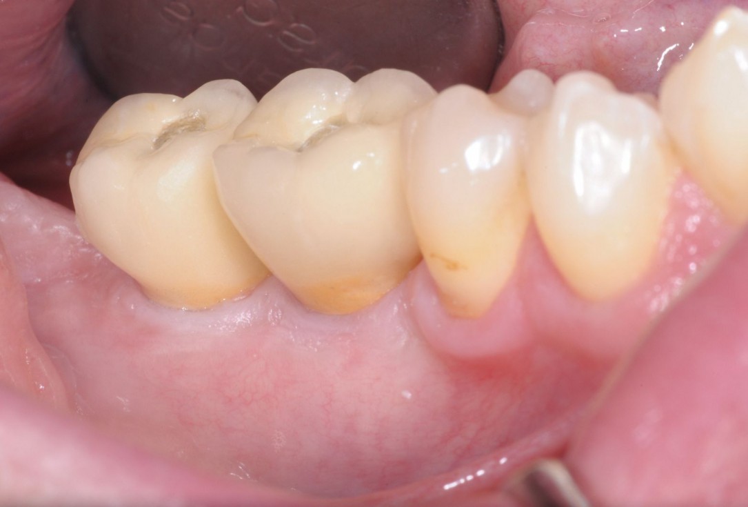
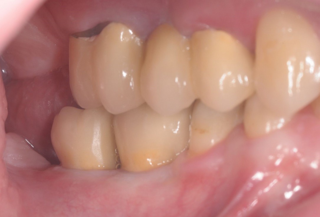
25/28 - Final prosthetic restauration with provisional screw channels - buccal viewThree-dimensional augmentation with maxgraft® cortico - Dr. R. Würdinger
-
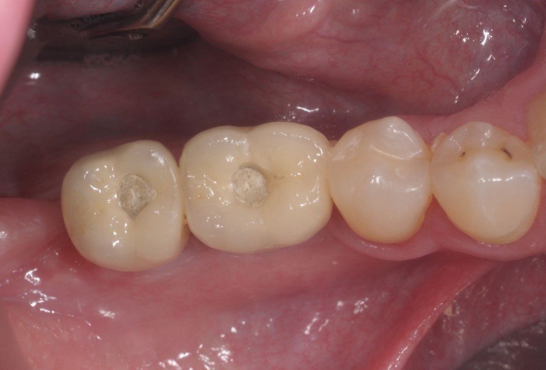
26/28 - Final prosthetic restauration with provisional screw channels - occlusal viewThree-dimensional augmentation with maxgraft® cortico - Dr. R. Würdinger
-
27/28 - Aesthetic and functional final resultThree-dimensional augmentation with maxgraft® cortico - Dr. R. Würdinger
-
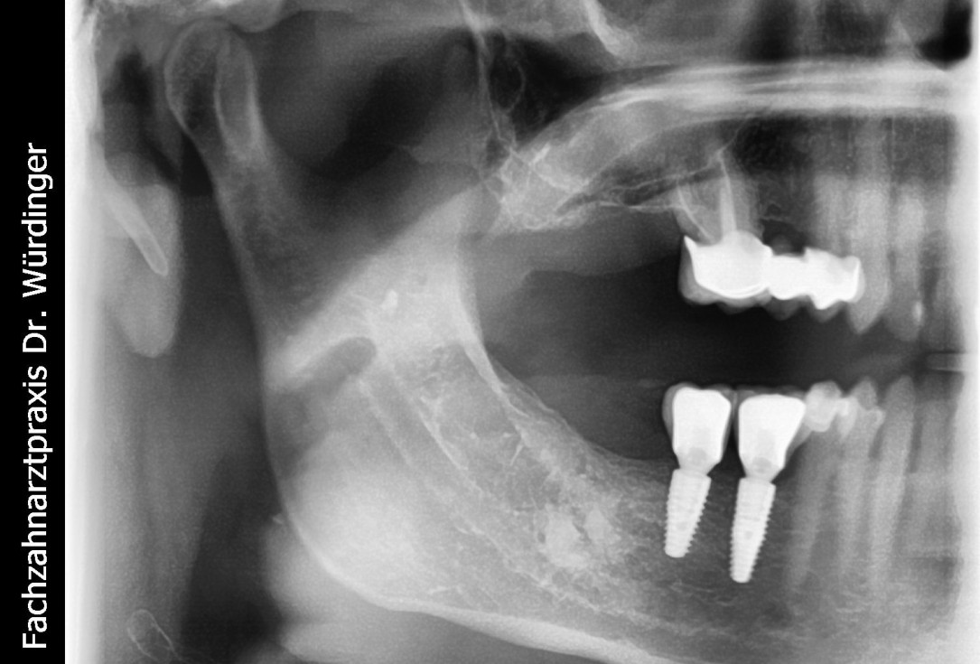
28/28 - Radiographic control of the final prosthesis - excellent stability of the augmented bone tissueThree-dimensional augmentation with maxgraft® cortico - Dr. R. Würdinger

Preparation of a single tooth defect with severely resorbed vestibular wall

Clinical situation

Occlusal view of attached maxgraft® cortico at the buccal site

Initial clinical situation – missing bonein regio 11, 12, 21, 22 and scarred soft tissue

OPG of the initial situation – provision of missing denture in regio 44 to 47 by a resin-retained bridge

Preoperative CBCT: vertical bone defects in the 3rd & 4th quadrant

Clinical situation

Severe atrophy of the ridge

Initial clinical situation
