GBR at implant site with collprotect®, cerabone® and Emdogain® - Dr. A. Nisio
-
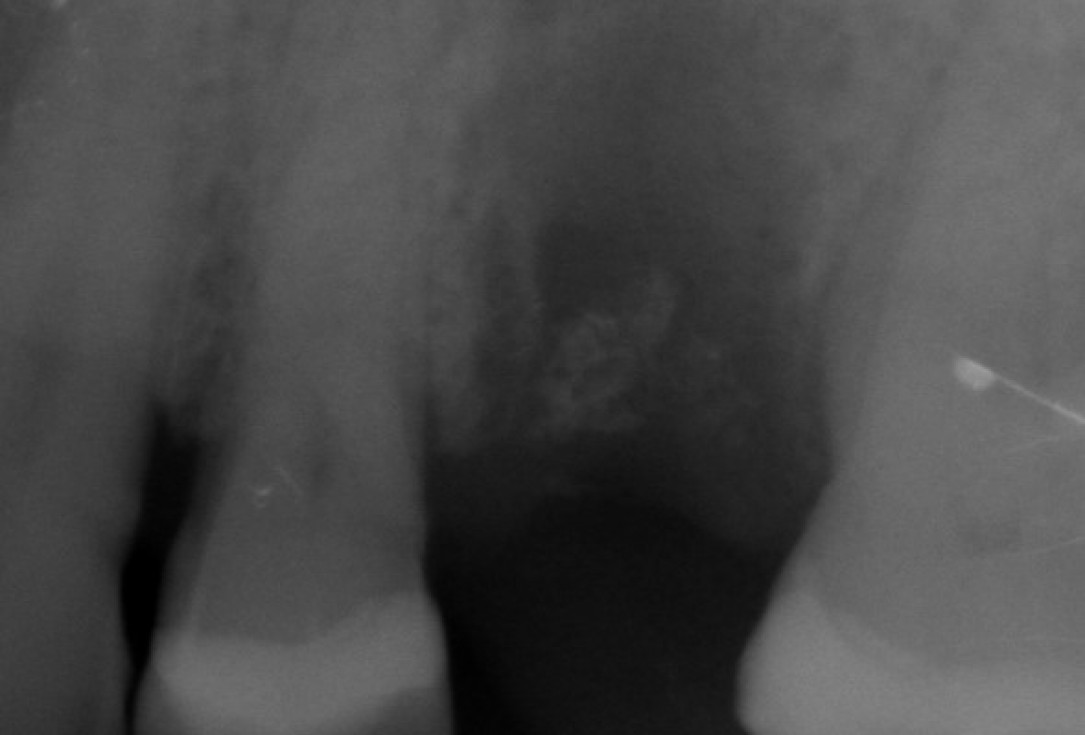
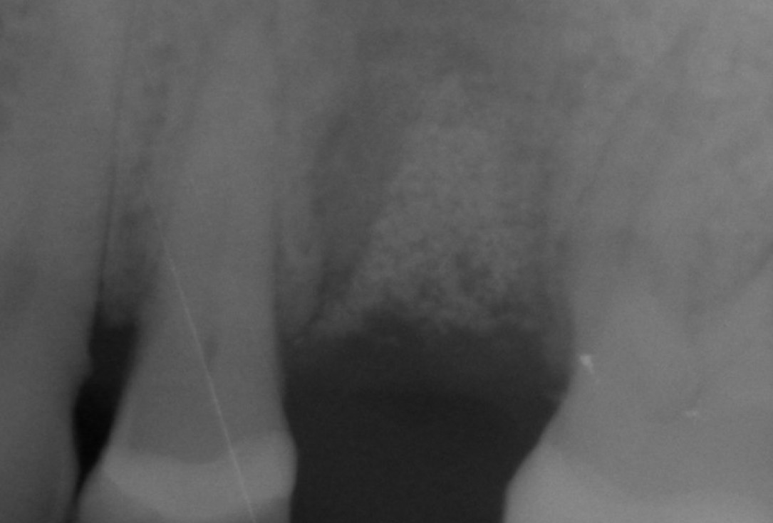
1/10 - Preoperative X-rayGBR at implant site with collprotect®, cerabone® and Emdogain® - Dr. A. Nisio
-
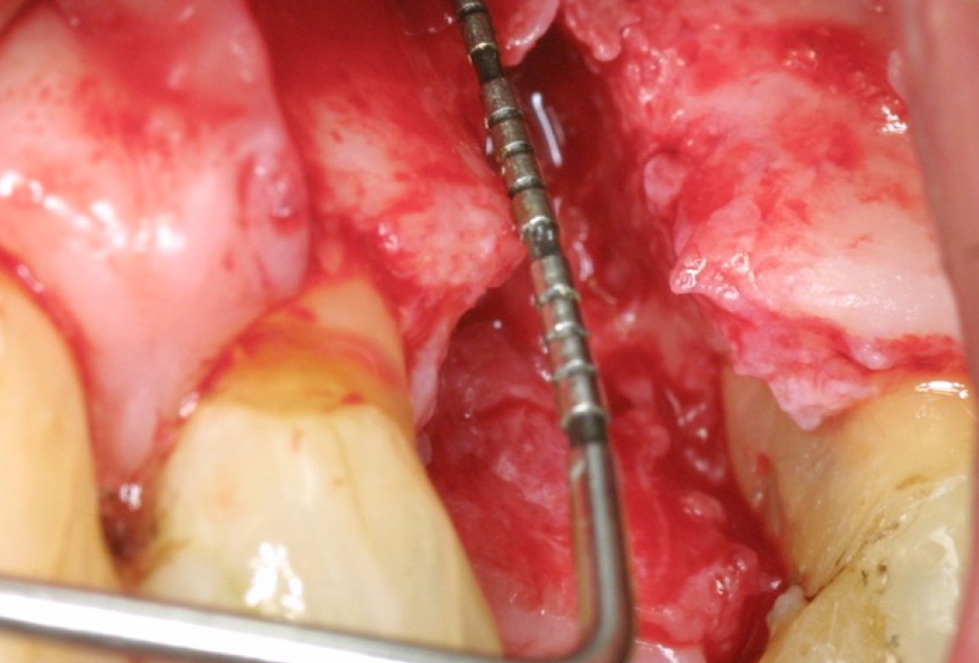
2/10 - Depth measurement of bony defect and root planing of adjacent teethGBR at implant site with collprotect®, cerabone® and Emdogain® - Dr. A. Nisio
-
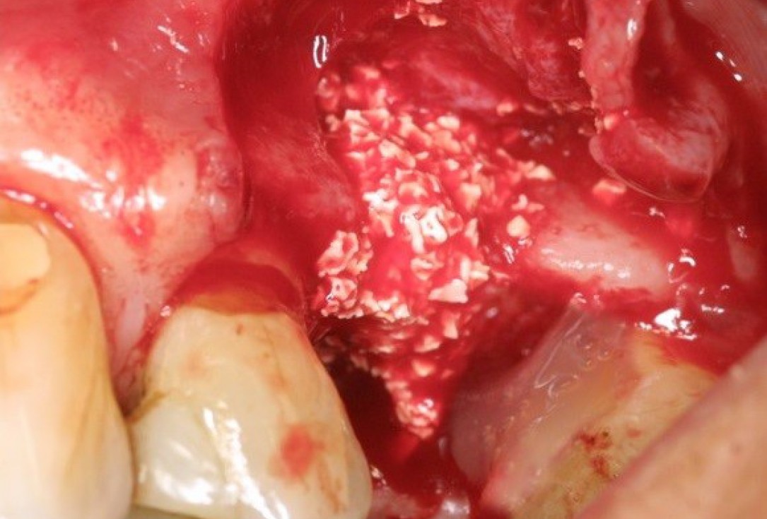
3/10 - Bone augmentation with cerabone® and treatment of root surface (tooth 26) with Emdogain®GBR at implant site with collprotect®, cerabone® and Emdogain® - Dr. A. Nisio
-
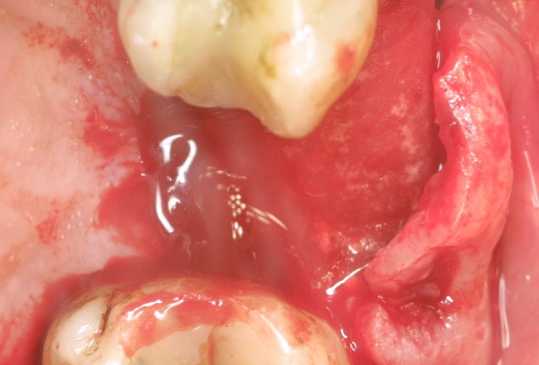
4/10 - Augmentation site covered with collprotect® membraneGBR at implant site with collprotect®, cerabone® and Emdogain® - Dr. A. Nisio
-
5/10 - X-ray at 3 monthsGBR at implant site with collprotect®, cerabone® and Emdogain® - Dr. A. Nisio
-
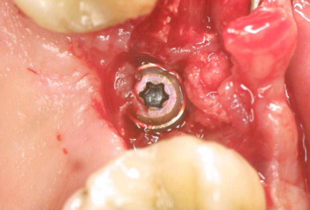
6/10 - Placement of Straumann bone level SLActive implant 3 months post-operativelyGBR at implant site with collprotect®, cerabone® and Emdogain® - Dr. A. Nisio
-
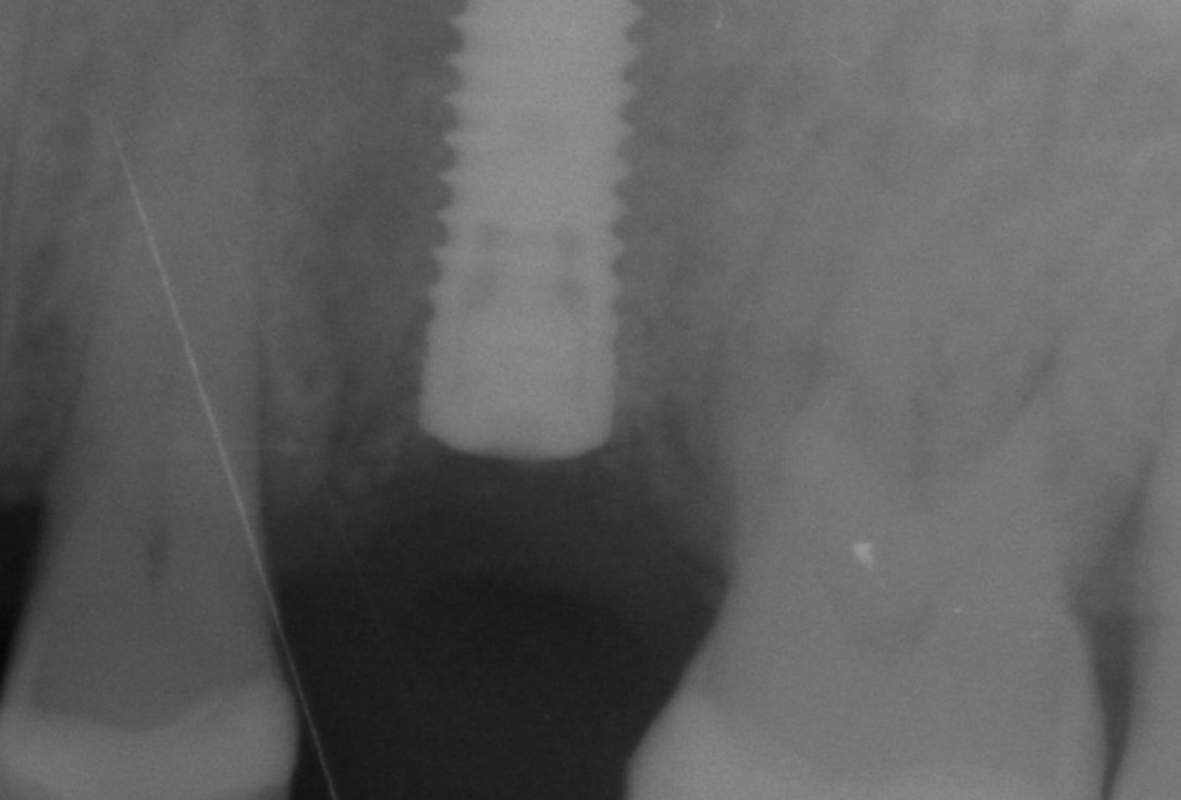
7/10 - Control X-ray 1 month after implant placementGBR at implant site with collprotect®, cerabone® and Emdogain® - Dr. A. Nisio
-
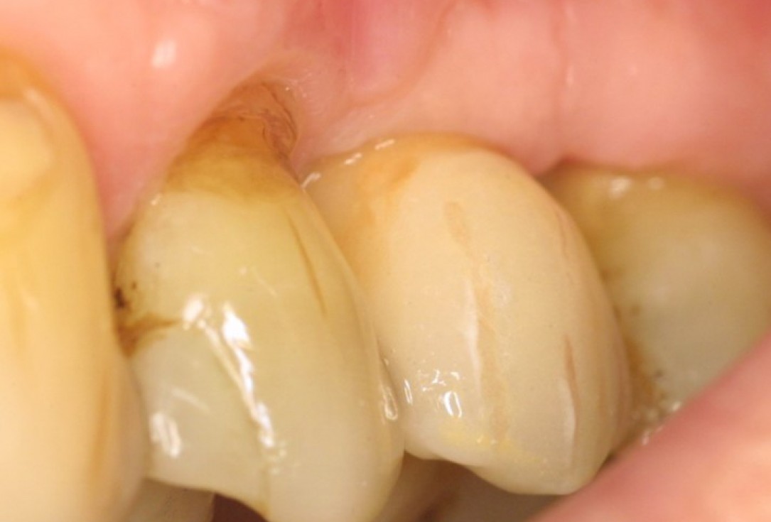
8/10 - After 1 year with final crownGBR at implant site with collprotect®, cerabone® and Emdogain® - Dr. A. Nisio
-
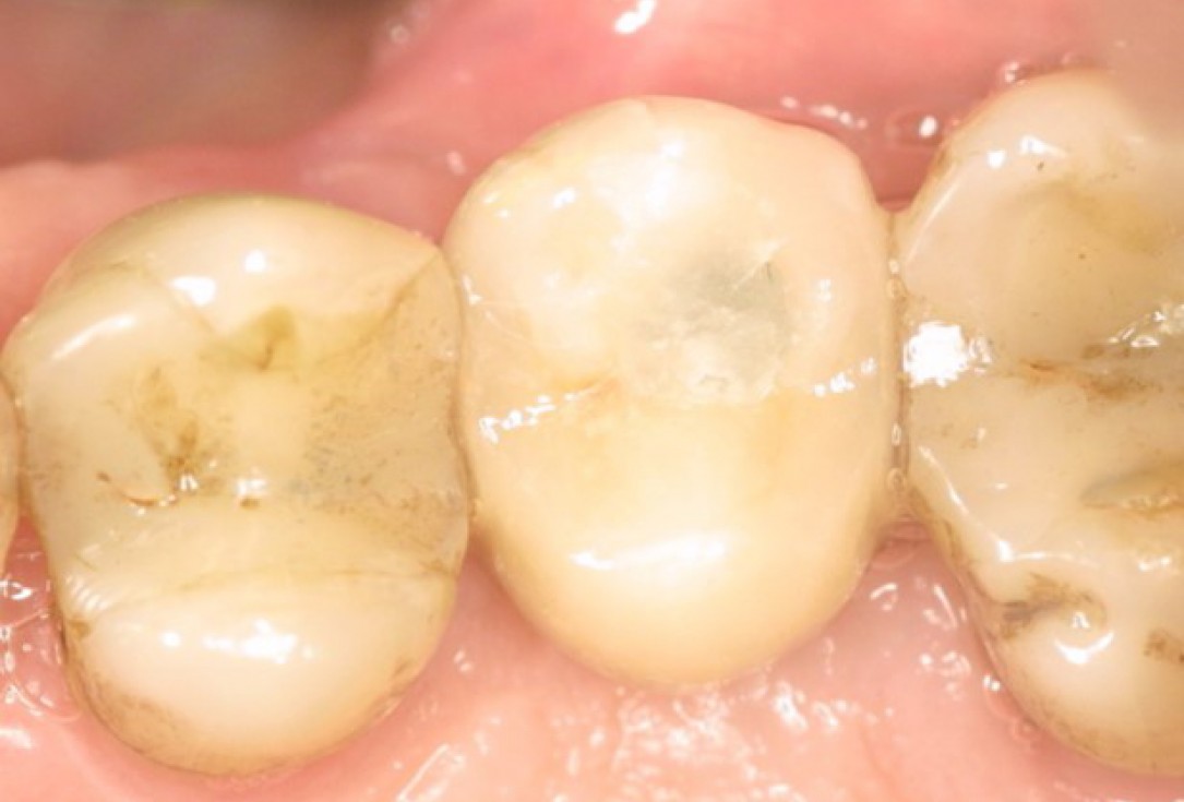
9/10 - Occlusal view of final crownGBR at implant site with collprotect®, cerabone® and Emdogain® - Dr. A. Nisio
-
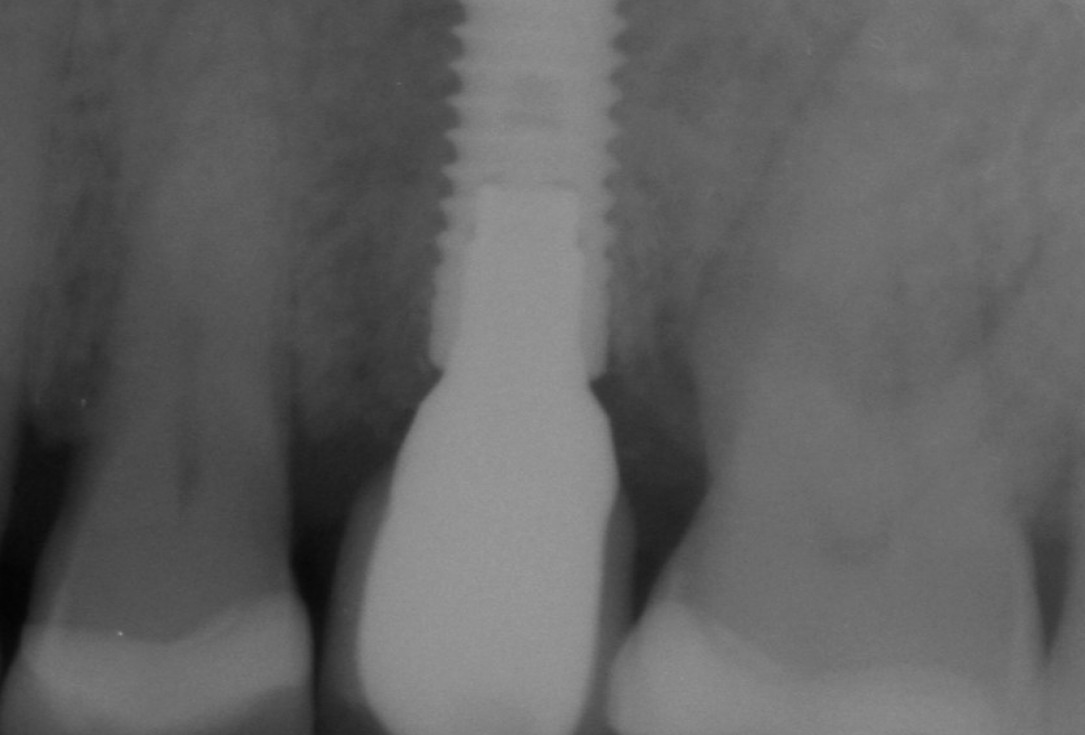
10/10 - X-ray 18 months post-operativelyGBR at implant site with collprotect®, cerabone® and Emdogain® - Dr. A. Nisio

Pre-operative OPG shows deep vertical intrabony defects on the distal aspects of teeth 13 and 14.

Pre-surgical probing reveals a deep intrabony defect on the distal aspect of the upper canine.

Pre-operative radiograph. Intrabony defect on the mesial aspect of tooth 14.

Initial X-ray presenting a very deep intrabony defect of tooth 21

Surgical presentation of the alveolar ridge with reduced amount of horizontal bone available

DVT control after sinusitis surgery, residual bone height 1 mm

OPG of the initial situation – provision of missing denture in regio 44 to 47 by a resin-retained bridge

The patient presented with a terminal fracture of the crown tooth number 12

Clinical situation with narrow alveolar ridge in the lower jaw

Extended horizontal and vertical defect of the maxilla following tumor resection and reconstruction with a scapula graft

Clinical situation of the edentulous distal maxilla before the surgery

DVT image showing the reduced amount of bone available in the area of the mental foramen
