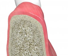
Socket seal technique

  • Socket seal technique with mucoderm®
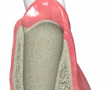
    Fresh extraction socket
  • Socket seal technique with mucoderm®
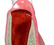
    Socket filled with a blood coagulum
  • Socket seal technique with mucoderm®
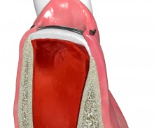
    Socket sealed with mucoderm®
  • Socket seal technique with mucoderm®
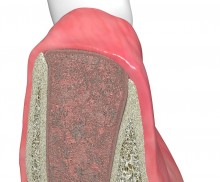
    Granulation tissue reorganization
  • Socket seal technique with mucoderm®
    Socket regeneration
According to the early implantation procedure, the implant is placed after tooth extraction, before the bony regeneration takes place within the socket. Typically, early implantation is performed about 4–8 weeks after tooth extraction; at this point, the healing of the soft tissue is complete and potential inflammations have disappeared. A closure of the soft tissue of the socket facilitates later augmentative procedures, if needed. Following extraction, the socket may be sealed with either a soft tissue transplant or a collagen matrix, such as mucoderm®, to stabilize the blood clot and protect it from bacterial contamination or physical damage. In the conventional approach, the socket is sealed with an autologous gingival transplant, which is harvested with a punch from the palate. Collagen matrix mucoderm® is a valid alternative to this approach that spares the patient the discomfort of the harvesting procedure.
Tooth extraction and socket sealing with mucoderm® - Dr. A. Rossi

Initial clinical situation showing strongly compromised tooth 21

Socket seal with mucoderm® punch

Intact socket following atraumatic tooth extraction

Post extraction socket sealing with mucoderm® - Dr. M. Frosecchi

X-ray showing endodontic failure of the molar

Rehydration and shaping of mucoderm®

Prior to application, mucoderm® should be rehydrated in a steril saline or blood for about 10 minutes. After rehydration, mucoderm® can be easily cut to shape with scissors or a scalpel.

De-epithlialization

To allow a migration of soft tissue cells from the surrounding tissue into the matrix, the ginigval margins should be de-epithelialized prior to application of mucoderm®.

Suturing of mucoderm®

To ensure a close contact to the marginal ginigiva, mucoderm® should be fixed with non-resorbable sutures, i.e., by single or double interrupted sutures.

Socket closed with mucoderm®

Upon tooth extraction, the alveolar socket fills with blood. The injury activates a coagulation cascade, which leads to the formation of a fibrin clot. This blood coagulum is the starting point of the healing and regeneration of the socket. Signaling factors in the blood promote blood vessel formation (angiogenesis); they also attract circulating progenitor and immune cells as well as connective tissue cells, which then migrate into the coagulum and form the granulation tissue. Within a few weeks, the granulation tissue is reorganized, and the osseous regeneration of the socket begins. Thus, the formation of a stable coagulum is of great importance for the regeneration of the socket; this can be achieved by sealing the socket.

The socket seal technique aims to protect the socket cavity and prevent a soft tissue collapse and shrinkage of the keratinized gingiva after extraction and prior to implantation. Following extraction, the socket may be sealed with either a soft tissue transplant or a collagen matrix that is sutured to the gingival margins of the socket. Alternatively, the socket may be filled with a bone graft material prior to sealing. The transplant supports the stabilization of the blood clot and protects it from bacterial contamination and physical damage.

For this particular indication, botiss has designed a circular mucoderm® matrix that can be easily applied during the socket seal technique and does not need further cutting.

Please Contact us for Literature.