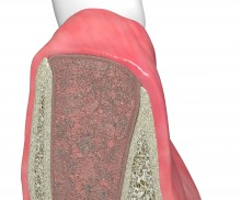
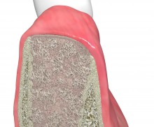
Natural healing

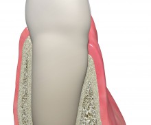
  • Natural healing - blood clot stabilization with collacone®
    Tooth planned for extraction
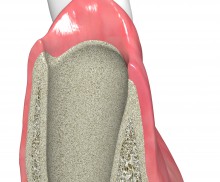
  • Natural healing - blood clot stabilization with collacone®
    Fresh extraction socket
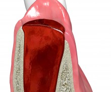
  • Natural healing - blood clot stabilization with collacone®
    Blood clot formed in the extraction socket
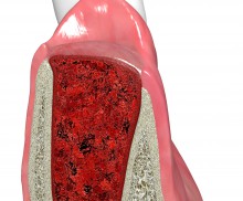
  • Natural healing - blood clot stabilization with collacone®
    collacone® stabilizing the blood clot
  • Natural healing - blood clot stabilization with collacone®
    Epithelial closure and ongoing regeneration of the socket
  • Natural healing - blood clot stabilization with collacone®
    Bony regeneration not completed at the time point of implantation
According to the early implantation procedure, the implant is placed after tooth extraction, before the bony regeneration takes place within the socket. Typically, an early implantation is performed about 4–8 weeks after tooth extraction; at this point, the healing of the soft tissue is complete and potential inflammations have disappeared. A closure of the soft tissue of the socket facilitates later augmentative procedures, if needed. The natural healing of the socket may be supported by applying a collagen sponge, such as collacone®—a hemostyptic natural porcine-collagen sponge. The application of collacone® into the socket supports the stabilization of the formed blood coagulum, while its form-fitted cone shape protects the wound area from food and bacteria.
Socket preservation with collacone® - Dr. K. Chmielewski

Central and lateral incisors are planned for extraction

Application of collacone®

Once in contact with a wet wound surface, collacone® sticks to the wound and forms a gel-like bond with blood. Fixation by cross- or holding sutures can increase the stability of the cone when applied in extraction sockets.

Rehydration of collacone®

A dry application of collacone® is recommended, because soaking or moistening of the cone prior to implantation may impair its hemostatic properties. At the defect site, the dry cone can rapidly soak up blood; during application, collacone® maintains its integrity also in the presence of blood.

contraindications for collacone®

As with most hemostatic agents, collagens are not intended for use in infected or contaminated wounds; in such a situation, they may actually act as a nidus for abscess formation and bacterial growth.

Upon tooth extraction, the alveolar socket fills with blood. The injury activates a coagulation cascade, which leads to the formation of a fibrin clot. This blood coagulum is the starting point of the healing and regeneration of the socket. Signaling factors in the blood promote blood vessel formation (angiogenesis); they also attract circulating progenitor and immune cells as well as connective tissue cells, which then migrate into the coagulum and form the granulation tissue. Within a few weeks, the granulation tissue is reorganized, and the osseous regeneration of the socket begins. Thus, the formation of a stable coagulum is of great importance for the regeneration of the socket; this can be achieved by sealing the socket.

Stabilization can either be achieved by sealing the socket with a soft tissue graft (socket seal technique) or by protecting it through the application of a collagen sponge (collacone®). The spongy structure of collacone® stabilizes the coagulum and provides an ideal structure for the adhesion of thrombocytes, fibroblasts, and osteoblasts. Fine blood vessels grow into and through the cone; as a result, the preliminary tissue formed in the alveoli is supplied with oxygen, nutrients, and the essential signaling molecules, which support its bony regeneration.

Although the bone volume is usually adequate after 4–8 weeks (since the resorption of the alveolar bone has not yet started), any existing bony defect of the alveoli may be treated at the moment of implantation by augmentation with a grafting material and barrier-membrane covering.

Please Contact us for Literature.