Broadening of the attached gingiva

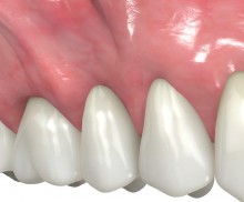
  • Extremely shallow vestibule and insufficient keratinized tissue
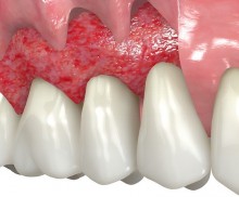
  • Split-thickness flap preparation
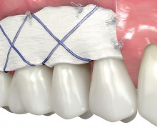
  • Fixation of mucoderm® to the periosteum
  • Regenerated soft tissue with broaden vestibule
The loss of the attached gingiva affects the oral health as well as the aesthetics and in addition compromises the natural support of the teeth or the dentures [1,2]. Vestibuloplasty is a pre-prosthetic surgical technique that has been developed to rebuild a sufficient vestibular depth by reducing the high muscle tensions that are streching to the maxillary or mandibular sides [3,4]. Two frequently used techniques to reconstruct deficient mucosal tissue are the free gingival graft (FGG) and the subepithelial connective tissue graft (SCTG) [5]. The gold standard, however, is still the autologous FGG from the hard palate especially when reconstructing a new band of attached gingiva [3,6,7]. Due to several limitations e.g. the amount of existing autologous tissue, discomfort during tissue harvesting and color mismatch [6], alternatives such as the mucoderm® acellular porcine matrix were developed.
Augmentation of the mandibular attached gingiva- Stricker

Initial clinical situation with loss of attached gingiva around the implants and shallow vestibule

Widening of the peri-implant keratinized mucosa-Horváth

Lack of sufficient keratinized mucosa following extensive horizontal ridge augmentation

botiss maxgraft® bonebuilder and vestibuloplasty with mucoderm® for ridge augmentation - Clinical case

Preoperative situation – Maxillary defect in area 14-16 (loss of implant 16 due to periimplantitis, tooth 14 extracted recently and area 15 already edentulous for a while)

botiss maxgraft® bonebuilder aesthetic reconstruction - Clinical case

Pre-operative clinical situation: changed color in the gingiva in the front maxilla

Initial situation

A minimum band of 1mm keratinized gingiva is required to enable regeneration of the acellular graft. The more native tissue surrounds the mucoderm® the better is the transformation of the biological information to the graft.

Rehydration

3-5 minute rehydration of mucoderm® in sterile saline solution or blood is essential prior to use. The flexibility of the mucoderm® can be adjusted by increasing the rehydration time.

Immobilization

A close contact between mucoderm® and the wound bed is required to guarantee revitalization of the grafted area.

Suturing

A close contact is crucial for uneventful healing and tissue integration and can be achieved by deep periosteal sling and superficial mattress sutures or single interrupted sutures. For a tension-free suturing of mucoderm® a sufficient depth of the vestibule bed is required.

Open-healing

mucoderm® can be left exposed for vestibuloplasty procedures without any additional wound dressings. Thereby, mucoderm® should be closely fixed to the periosteum.

After-care

The operating field must be treated with care after surgery and patients should be recalled weekly to evaluate the healing.

mucoderm® for broadening of the attached gingiva
mucoderm® zur Verbreiterung der befestigten Gingiva

mucoderm® not only ensures an ideal color match with the surrounding tissue, but also reduces pain and discomfort of the patient. After a short rehydration in sterile saline solution or blood, the matrix can be easily trimmed. Following the preparation of a split-thickness flap, the mobile mucosa is positioned apically and fixed to the deep vestibular fold, which allows a new vestibule to be formed. The mucoderm® should be placed in close contact to the exposed periosteum and securely fixed to allow its revitalization by underlying cells and blood vessels. Please note that the open-healing will likely reduce the degradation time of mucoderm®.

[1] Arnoux et al. Int J Oral Maxillofac. Implants. 1998; 13(4): 565-8
[2] George and Dhir. J Int Clin Dent Res Organ 2015; 7 (1):119-31
[3] Cranin AN. J Oral Implantol. 2002; 28(5): 230-7
[4] Heller et al. J Oral Implantol 2000; 26(2):91-103
[5] OH SL. Gen Dent. 2009; 57(4): 381-5
[6] Fröschl and Kerscher. J Craniomaxillofac Surg. 1997; 25(2): 85-90
[7] Edel A. Periodontol Clin Investig. 1998; 20:12-20