Bone regeneration and implant-supported rehabilitation of a periodontally involved incisor - Dr. A. Eslava Vanegas
-
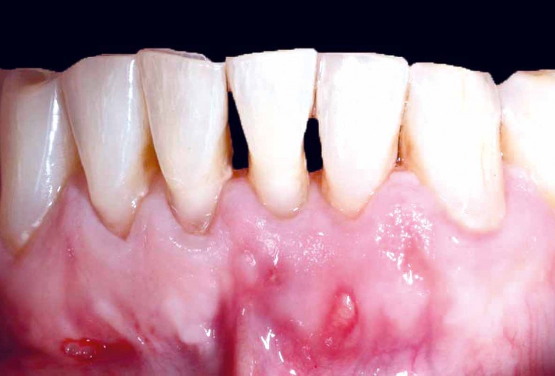
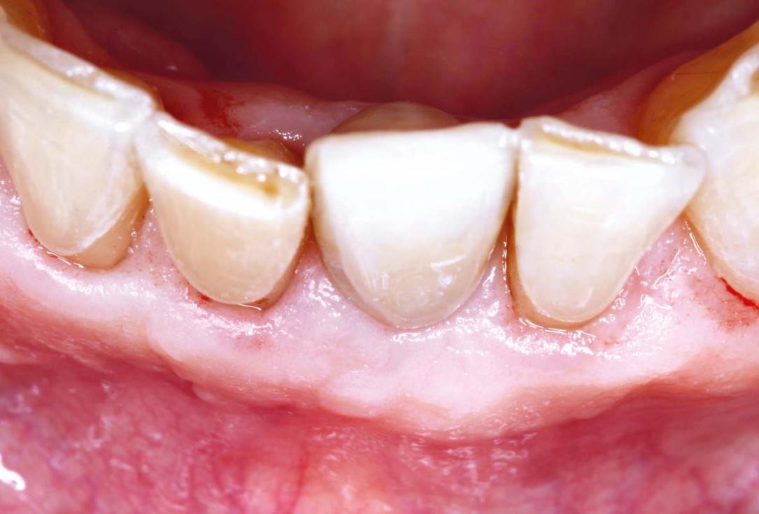
01/22 - Initial clinical situationBone regeneration and implant-supported rehabilitation of a periodontally involved incisor - Dr. A. Eslava Vanegas
-
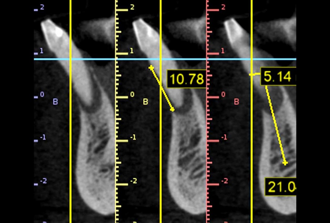
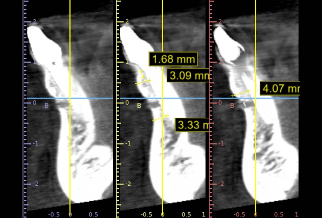
02/22 - Initial CT scan showing 10 mm bone lossBone regeneration and implant-supported rehabilitation of a periodontally involved incisor - Dr. A. Eslava Vanegas
-
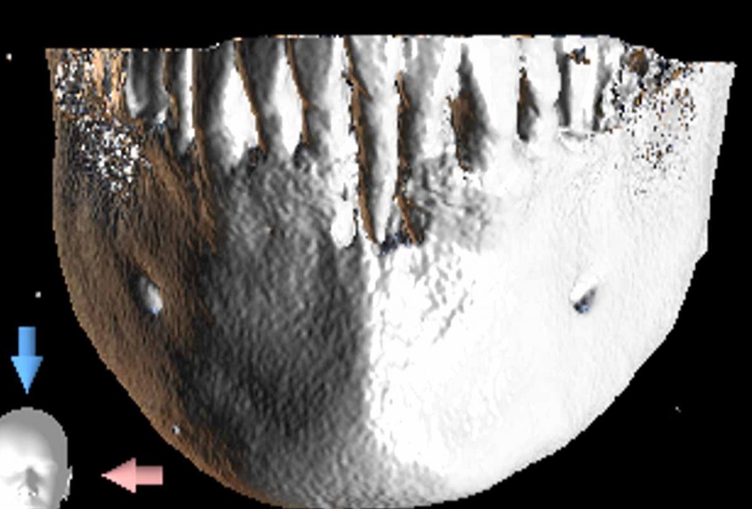
03/22 - Initial 3D bone reconstructionBone regeneration and implant-supported rehabilitation of a periodontally involved incisor - Dr. A. Eslava Vanegas
-
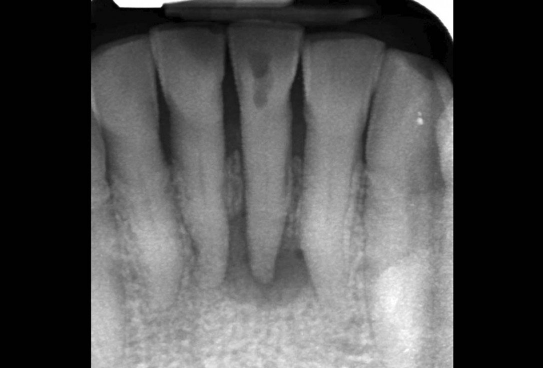
04/22 - Endo-perio lesion at tooth 31Bone regeneration and implant-supported rehabilitation of a periodontally involved incisor - Dr. A. Eslava Vanegas
-
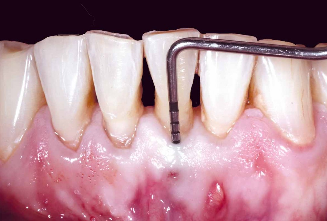
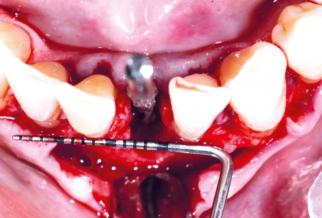
05/22 - 12 mm probing depthBone regeneration and implant-supported rehabilitation of a periodontally involved incisor - Dr. A. Eslava Vanegas
-
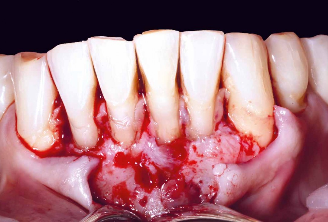
06/22 - After raising a mucoperiosteal flap with granulation tissue at the apexBone regeneration and implant-supported rehabilitation of a periodontally involved incisor - Dr. A. Eslava Vanegas
-
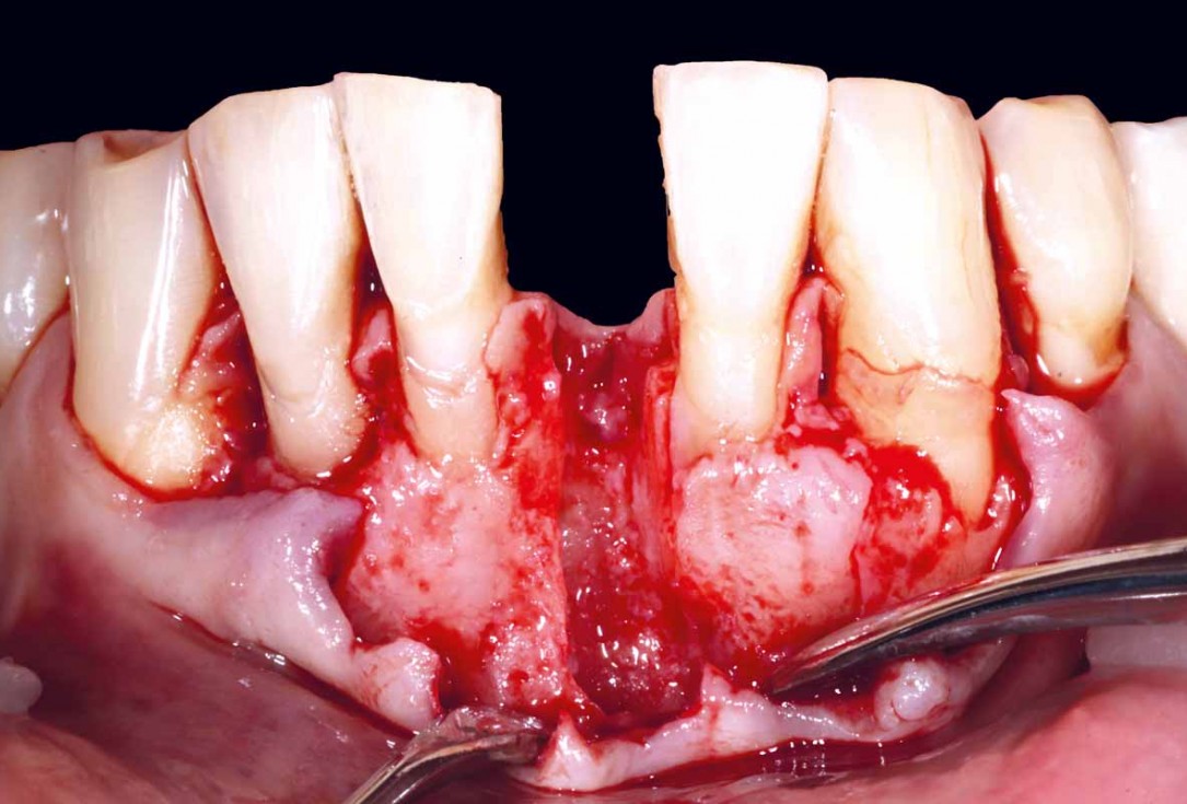
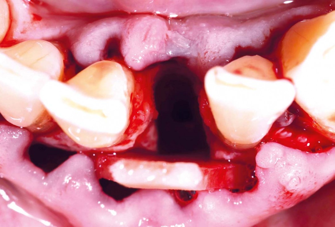
07/22 - After tooth extraction, total buccal bone loss visibleBone regeneration and implant-supported rehabilitation of a periodontally involved incisor - Dr. A. Eslava Vanegas
-
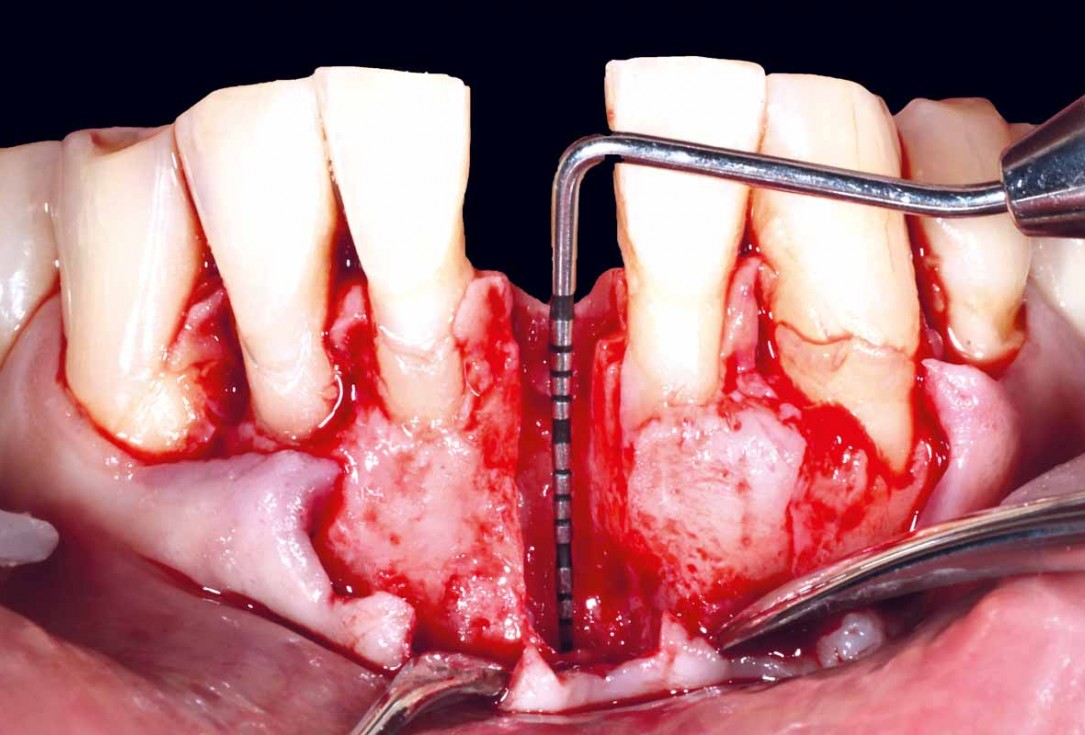
08/22 - 10 mm buccal bone lossBone regeneration and implant-supported rehabilitation of a periodontally involved incisor - Dr. A. Eslava Vanegas
-
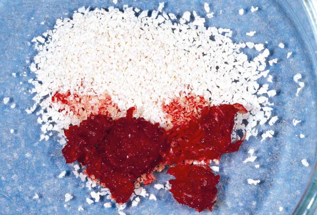
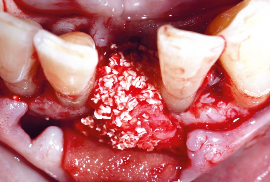
09/22 - Autologous chips with cerabone® granules in preparation for bone graftingBone regeneration and implant-supported rehabilitation of a periodontally involved incisor - Dr. A. Eslava Vanegas
-
10/22 - Checking future 3D implant positionBone regeneration and implant-supported rehabilitation of a periodontally involved incisor - Dr. A. Eslava Vanegas
-
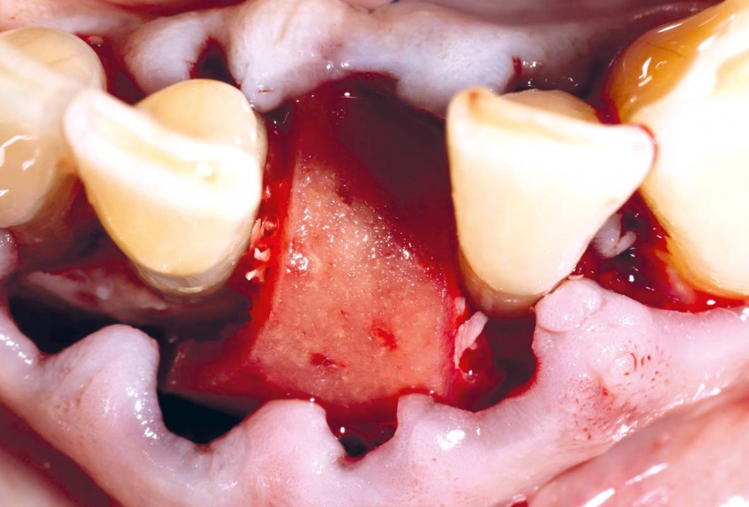
11/22 - Pre-hydrated mucoderm® in placeBone regeneration and implant-supported rehabilitation of a periodontally involved incisor - Dr. A. Eslava Vanegas
-
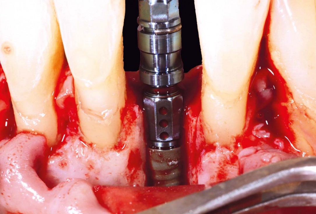
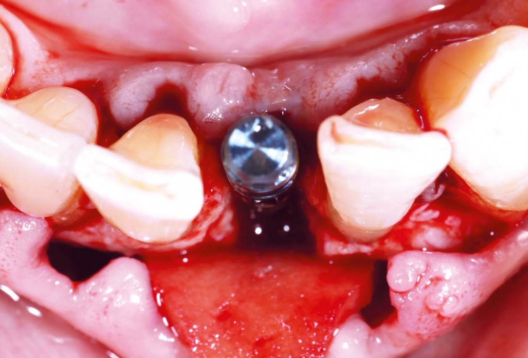
12/22 - 2.9 mm BLT Straumann SLActive implant insertedBone regeneration and implant-supported rehabilitation of a periodontally involved incisor - Dr. A. Eslava Vanegas
-
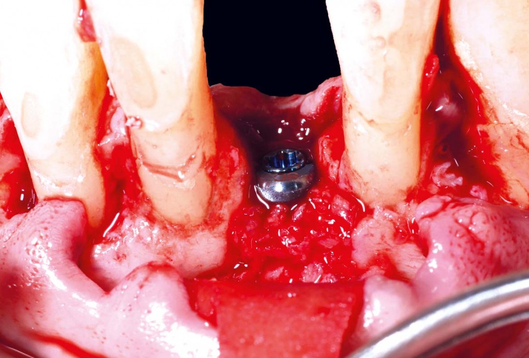
13/22 - Checking 3D implant positionBone regeneration and implant-supported rehabilitation of a periodontally involved incisor - Dr. A. Eslava Vanegas
-
14/22 - 2 mm healing abutment inserted. Autologous bone chips placed on the implant surfaceBone regeneration and implant-supported rehabilitation of a periodontally involved incisor - Dr. A. Eslava Vanegas
-
15/22 - Second layer of autograft mixed with cerabone and PRF appliedBone regeneration and implant-supported rehabilitation of a periodontally involved incisor - Dr. A. Eslava Vanegas
-
16/22 - mucoderm® covering the bone defect, acting as a barrier membraneBone regeneration and implant-supported rehabilitation of a periodontally involved incisor - Dr. A. Eslava Vanegas
-
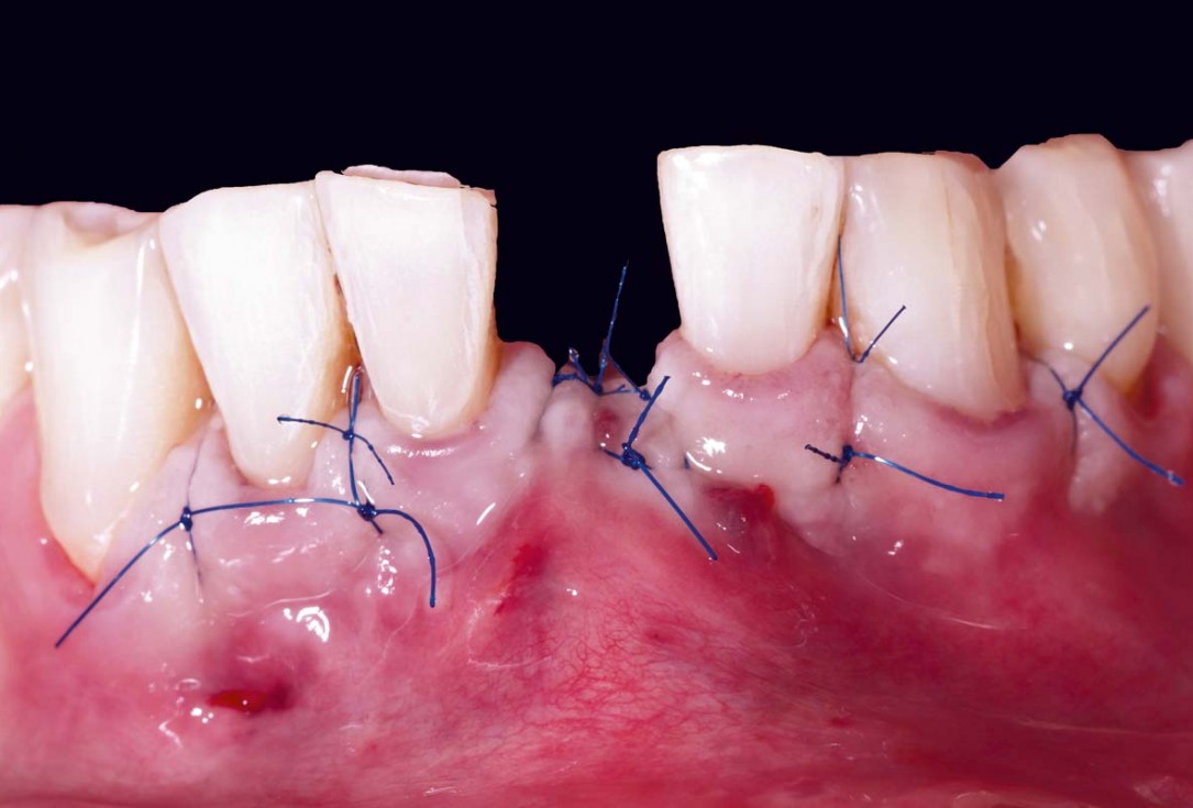
17/22 - Flap closureBone regeneration and implant-supported rehabilitation of a periodontally involved incisor - Dr. A. Eslava Vanegas
-
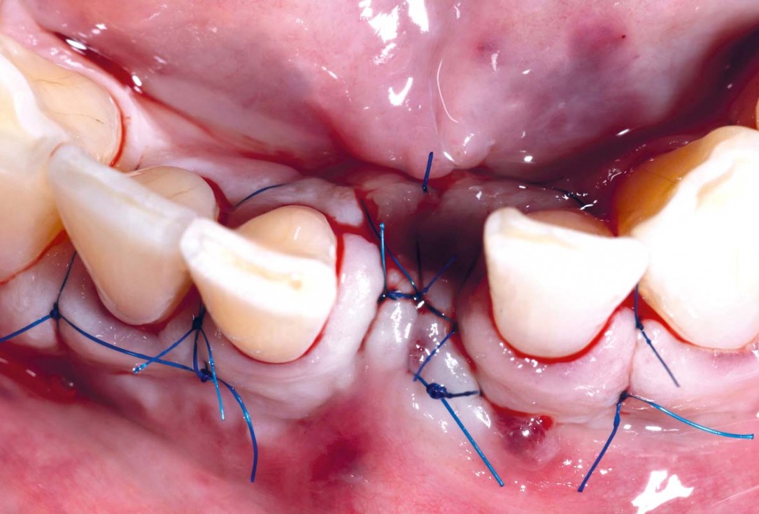
18/22 - Occlusal viewBone regeneration and implant-supported rehabilitation of a periodontally involved incisor - Dr. A. Eslava Vanegas
-
19/22 - Situation 8 months post-operativeBone regeneration and implant-supported rehabilitation of a periodontally involved incisor - Dr. A. Eslava Vanegas
-
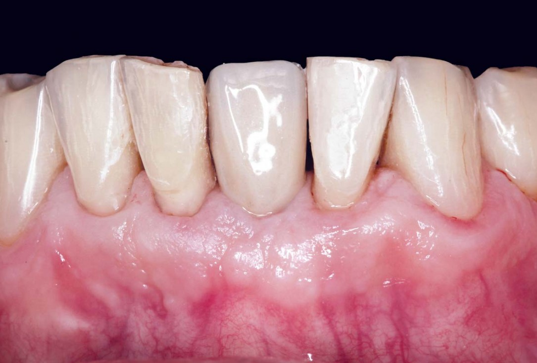
20/22 - Final prosthetic restoration 3 years post-surgeryBone regeneration and implant-supported rehabilitation of a periodontally involved incisor - Dr. A. Eslava Vanegas
-
21/22 - Occlusal viewBone regeneration and implant-supported rehabilitation of a periodontally involved incisor - Dr. A. Eslava Vanegas
-
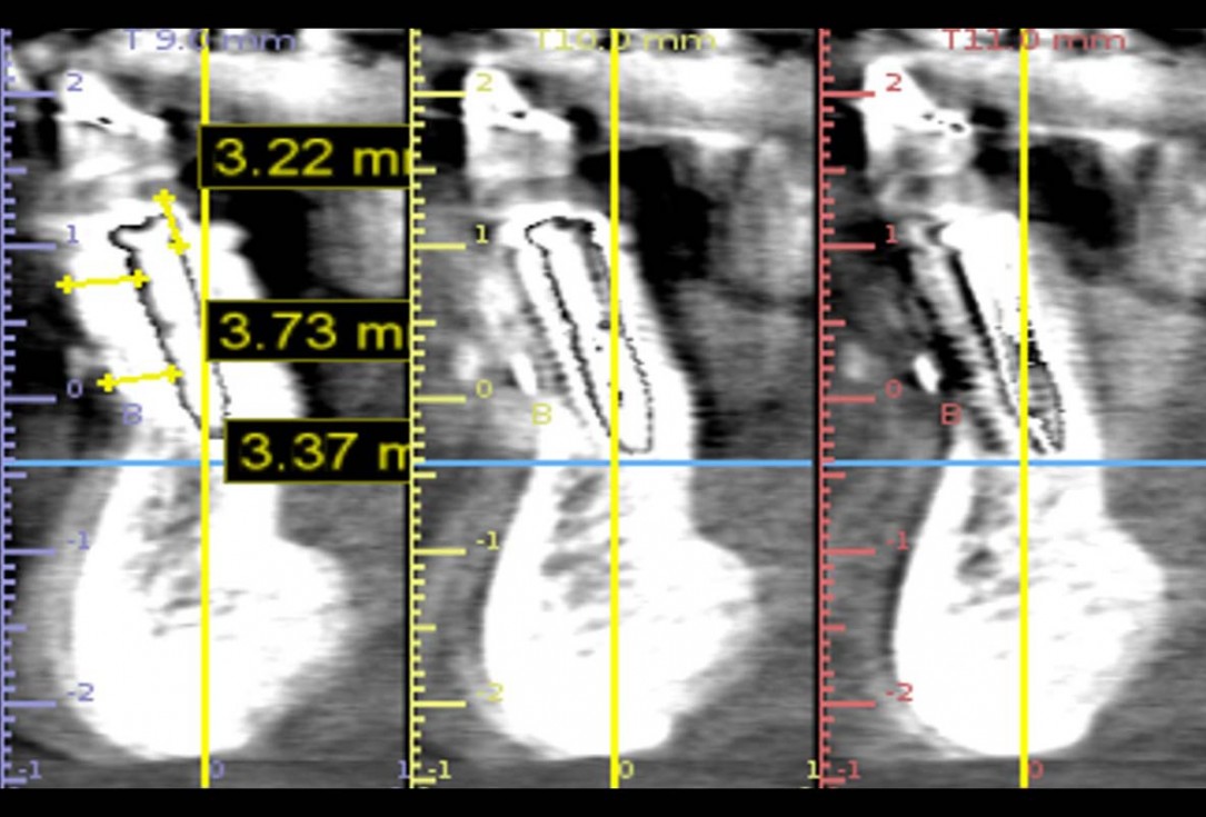
22/22 - Final CT scan 3 years post-operativeBone regeneration and implant-supported rehabilitation of a periodontally involved incisor - Dr. A. Eslava Vanegas

Initial Orthopantomograph X-Ray

Clinical situation before extraction and implantation

X-ray control before tooth extraction

Clinical view of the case.

Pre-operative x-ray image, teeth 43, 44, 45, 46 and 47 planned for extraction

The patient presented with a terminal fracture of the crown tooth number 12

47 years old patient referred by another dentist after suffering a fall while fishing

Preoperative Ortopantomogram of the teeth planned for extraction

Initial situation pre-op: Central incisors with mobility 3

Pre-operative OPG, tooth 36 planned for extraction

Initial situation with fractured central incisors

Initial clinical situation - Central incisors with dental destruction and periapical pathology

Immediately placed implant covered with permamem®. permamem® passively immobilized by sutures and intentionally left exposed to the oral cavity.

Pre-operative situation showing tooth 21 with deep periodontal pocket. Tooth presented with mobility grade III.

Initial view of the case. Discoloration of 1.1 and mild class I gingival recession

Pre-operative OPG, teeth 24, 25, and 26 planned for extraction

Initial situation with broken tooth 46
