Bone augmentation in aesthetic zone with maxgraft® bonering - Dr. A. Patel
-
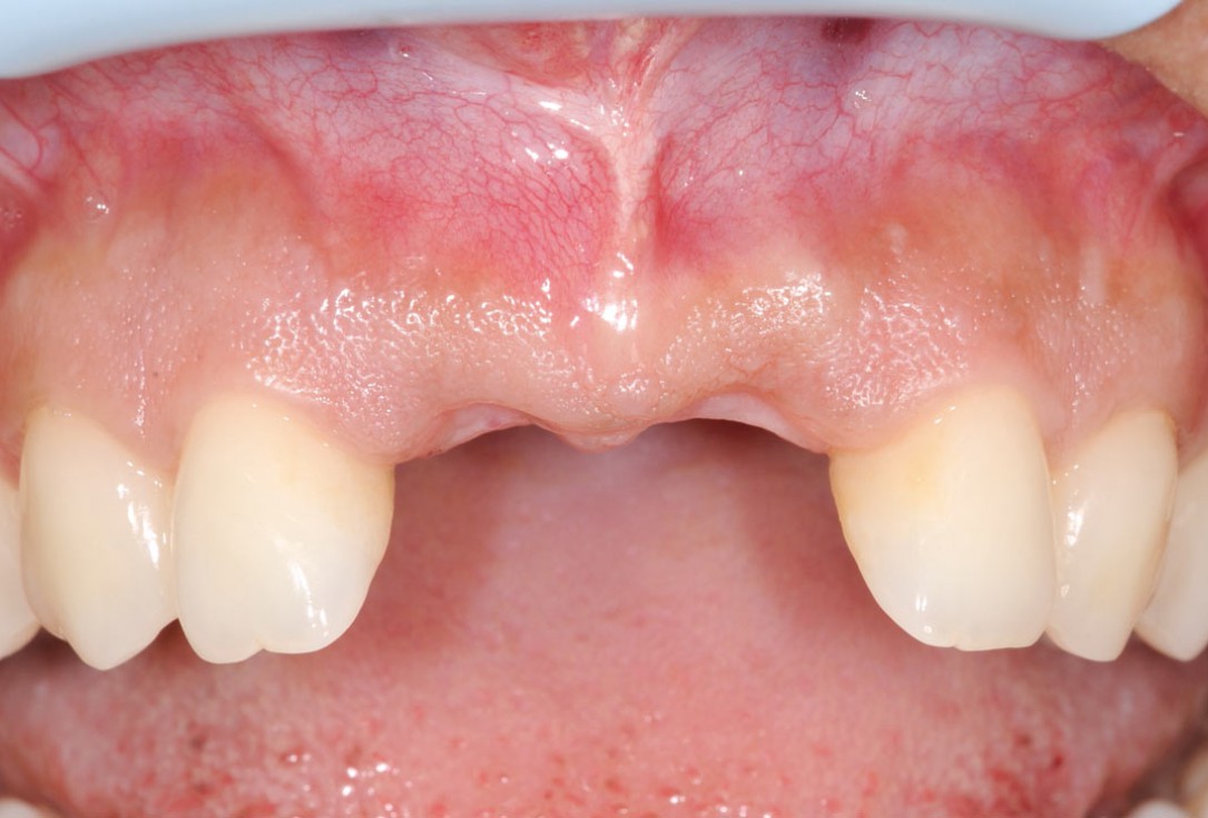
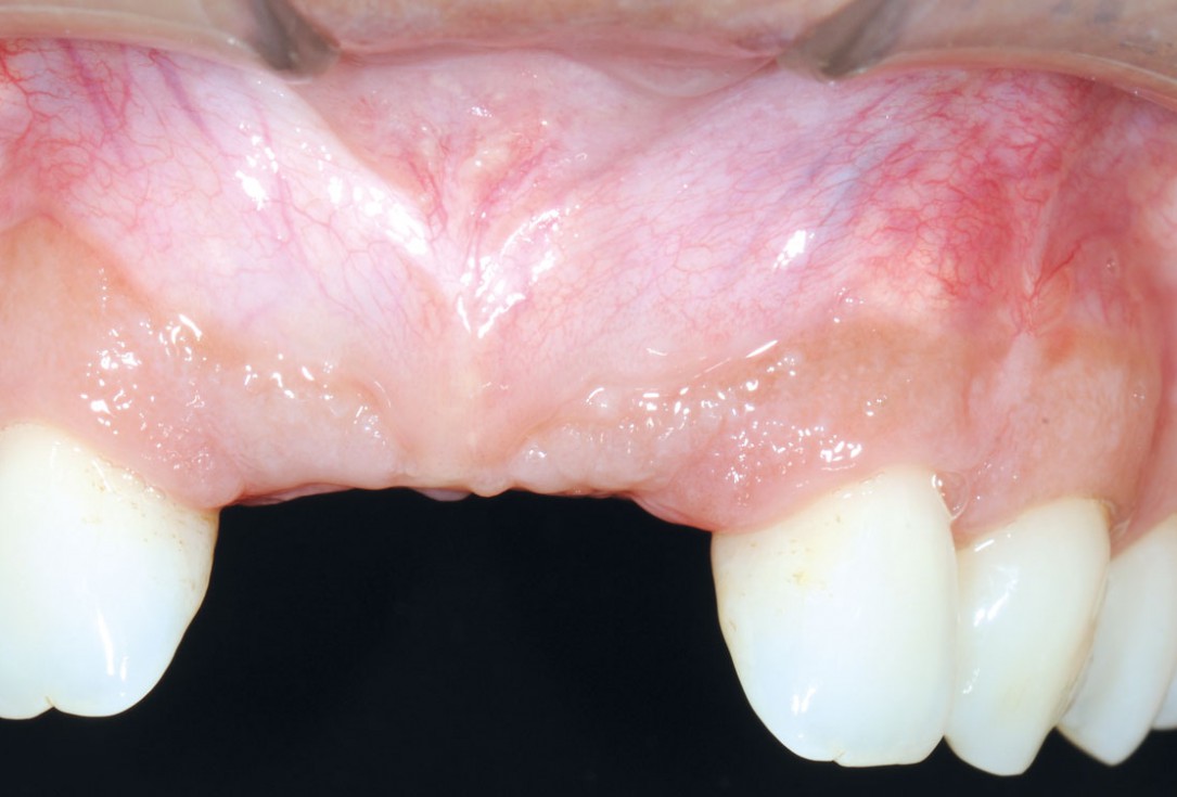
1/26 - Initially bridge retained incisorsBone augmentation in aesthetic zone with maxgraft® bonering - Dr. A. Patel
-
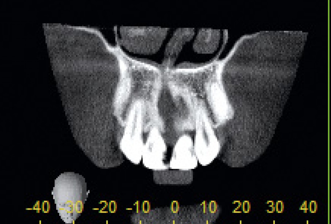
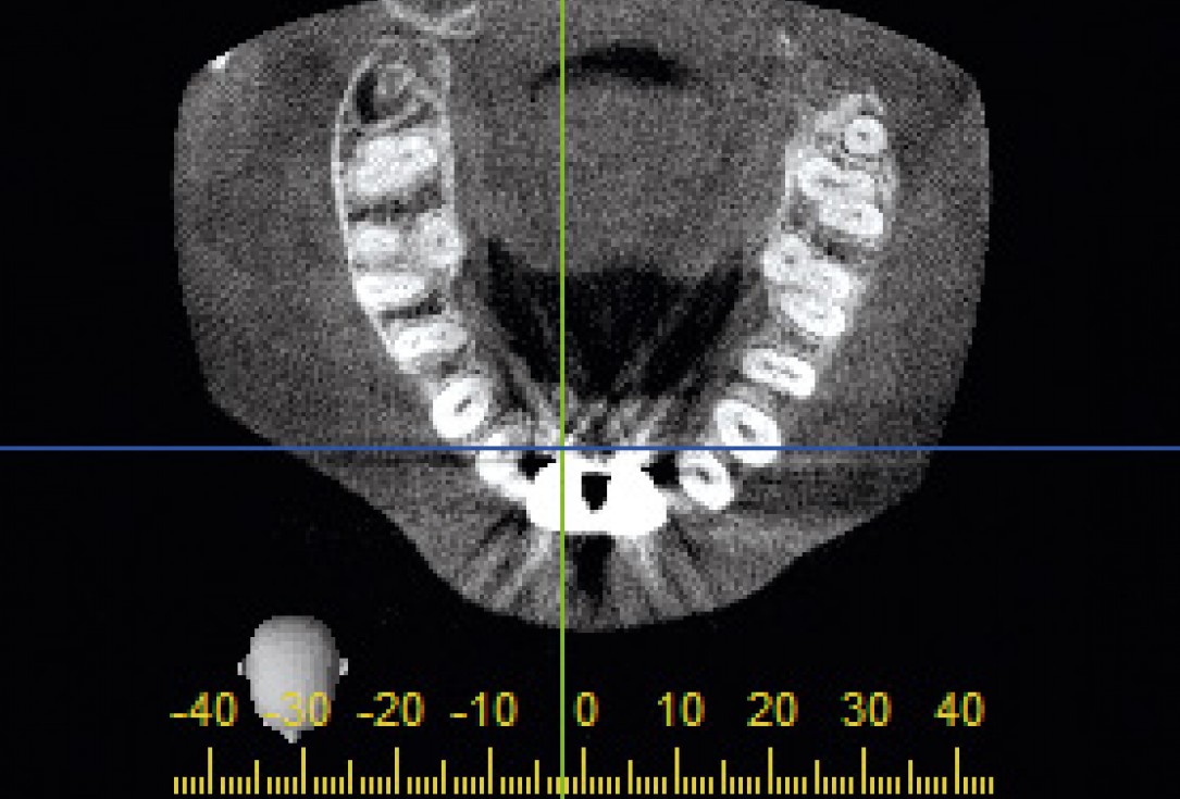
2/26 - CT scan reveals major bone loss in frontal maxillaBone augmentation in aesthetic zone with maxgraft® bonering - Dr. A. Patel
-
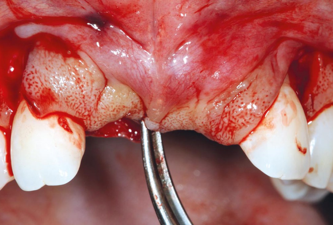
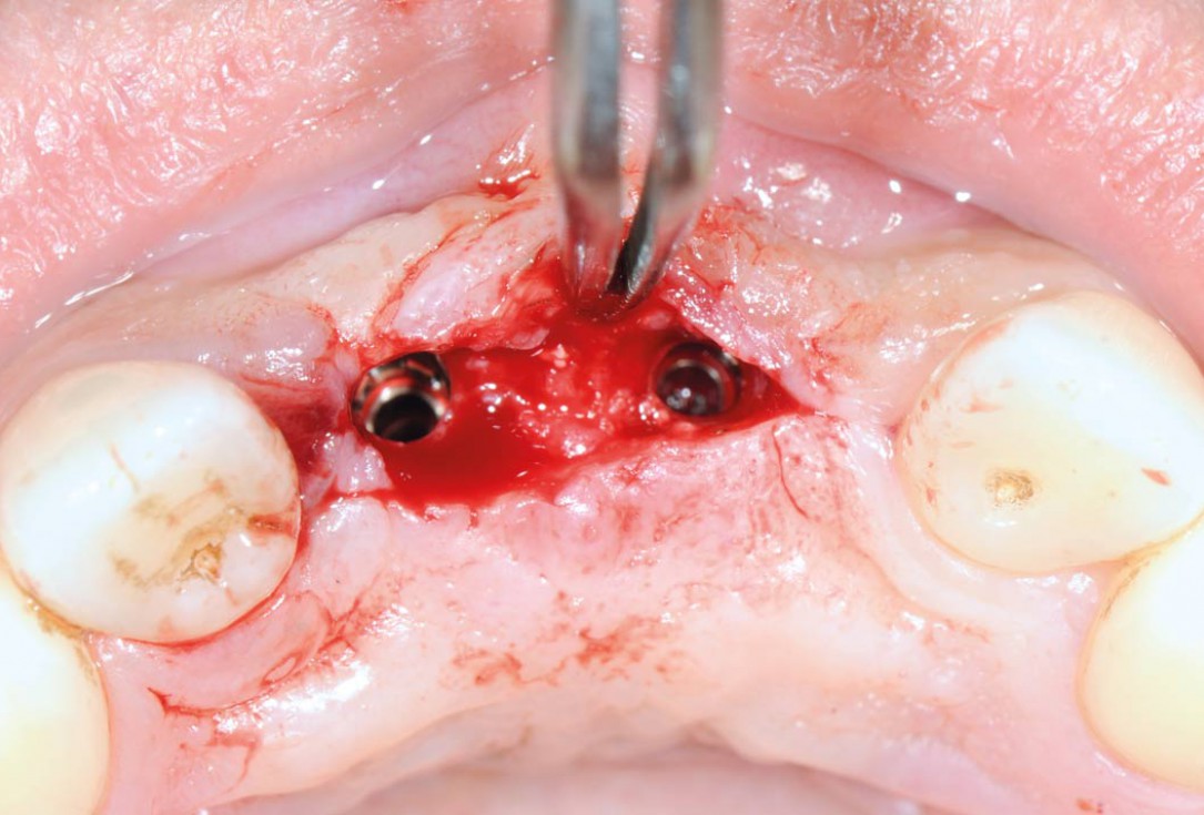
3/26 - Big bone defect visible after opening the flapBone augmentation in aesthetic zone with maxgraft® bonering - Dr. A. Patel
-
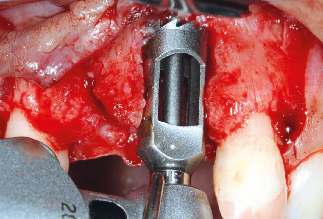
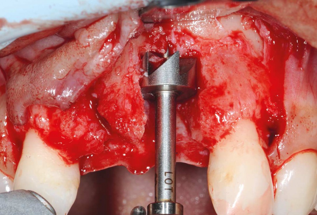
4/26 - Determine the defect size with the trephineBone augmentation in aesthetic zone with maxgraft® bonering - Dr. A. Patel
-
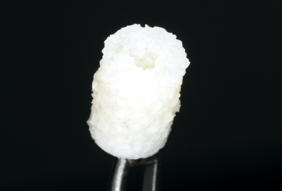
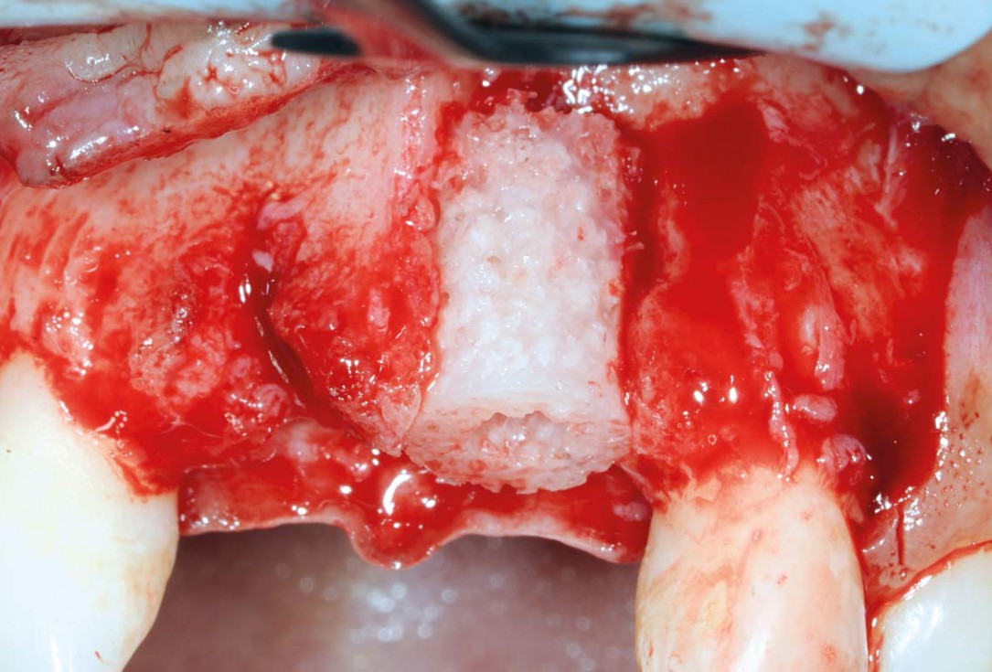
5/26 - Hydrated maxgraft® bonering 7 mmBone augmentation in aesthetic zone with maxgraft® bonering - Dr. A. Patel
-
6/26 - Preparing the site for maxgraft® boneringBone augmentation in aesthetic zone with maxgraft® bonering - Dr. A. Patel
-
7/26 - Paving the surface of the recipient site to create press-fitting of the ringBone augmentation in aesthetic zone with maxgraft® bonering - Dr. A. Patel
-
8/26 - Placement of maxgraft® bonering 10 mm heightBone augmentation in aesthetic zone with maxgraft® bonering - Dr. A. Patel
-
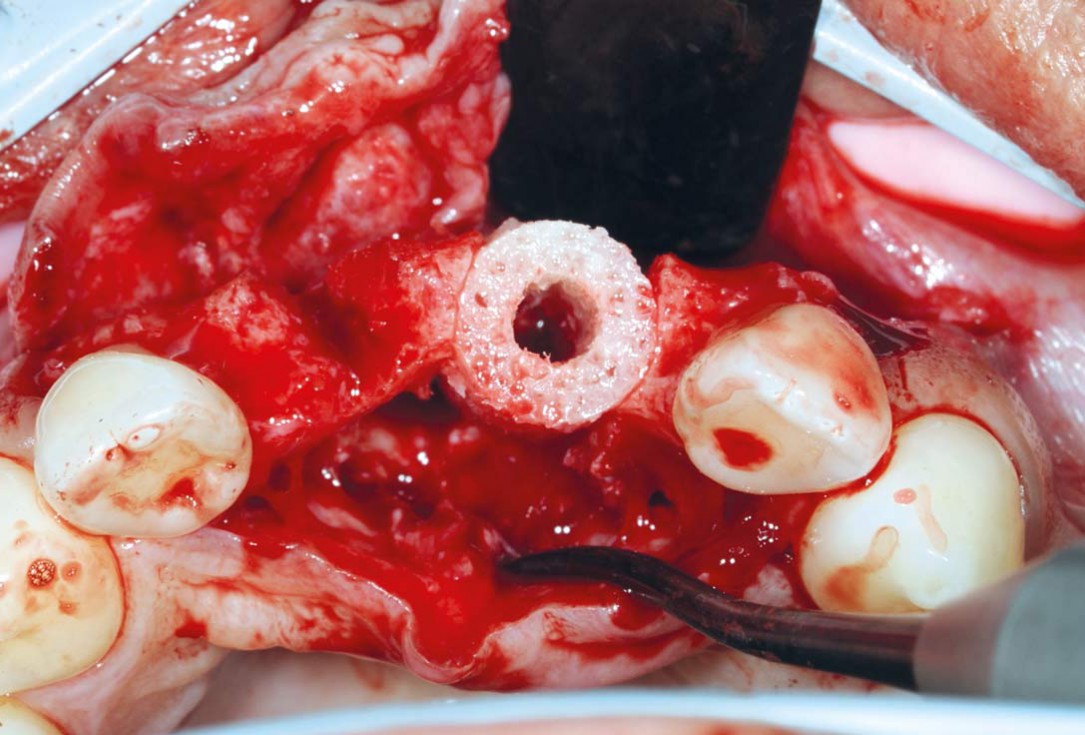
9/26 - Occlusal view after placing the bone graft confirms ideal positioningBone augmentation in aesthetic zone with maxgraft® bonering - Dr. A. Patel
-
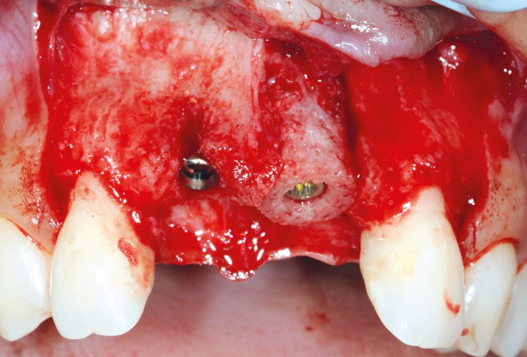
10/26 - Fixation of maxgraft® bonering with a Straumann® SLActive Bone Level Tapered ImplantBone augmentation in aesthetic zone with maxgraft® bonering - Dr. A. Patel
-
11/26 - Implantation in region 11Bone augmentation in aesthetic zone with maxgraft® bonering - Dr. A. Patel
-
12/26 - Mobilizing the flapBone augmentation in aesthetic zone with maxgraft® bonering - Dr. A. Patel
-
13/26 - Covering the site with cerabone® to prevent resorptionBone augmentation in aesthetic zone with maxgraft® bonering - Dr. A. Patel
-
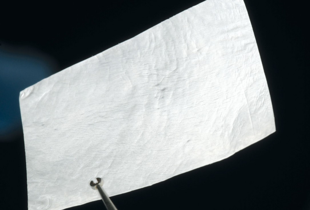
14/26 - Jason® membraneBone augmentation in aesthetic zone with maxgraft® bonering - Dr. A. Patel
-
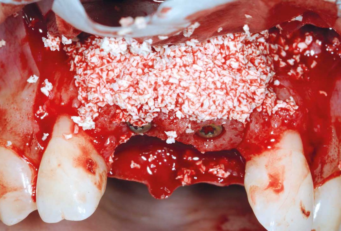
15/26 - Adding more cerabone® after attaching the Jason® membraneBone augmentation in aesthetic zone with maxgraft® bonering - Dr. A. Patel
-
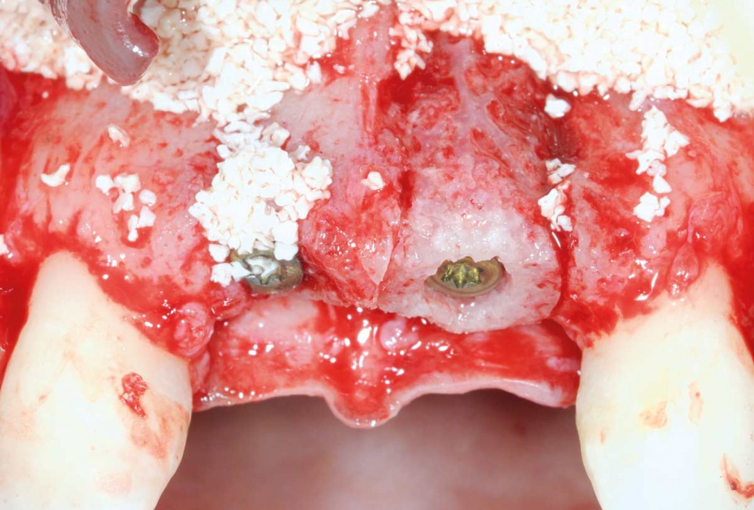
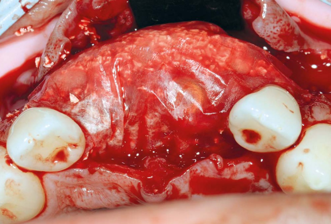
16/26 - Bone volume gained buccally and site covered with Jason® membraneBone augmentation in aesthetic zone with maxgraft® bonering - Dr. A. Patel
-
17/26 - Mattress sutures to stabilize the graftBone augmentation in aesthetic zone with maxgraft® bonering - Dr. A. Patel
-
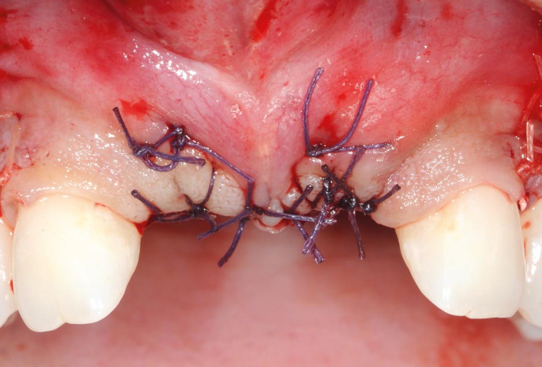
18/26 - Sutured free of tension with vycrilBone augmentation in aesthetic zone with maxgraft® bonering - Dr. A. Patel
-
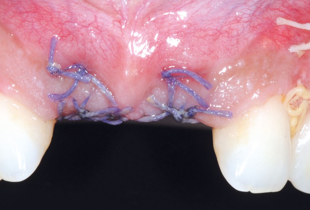
19/26 - 3 weeks post-op: eventless healingBone augmentation in aesthetic zone with maxgraft® bonering - Dr. A. Patel
-
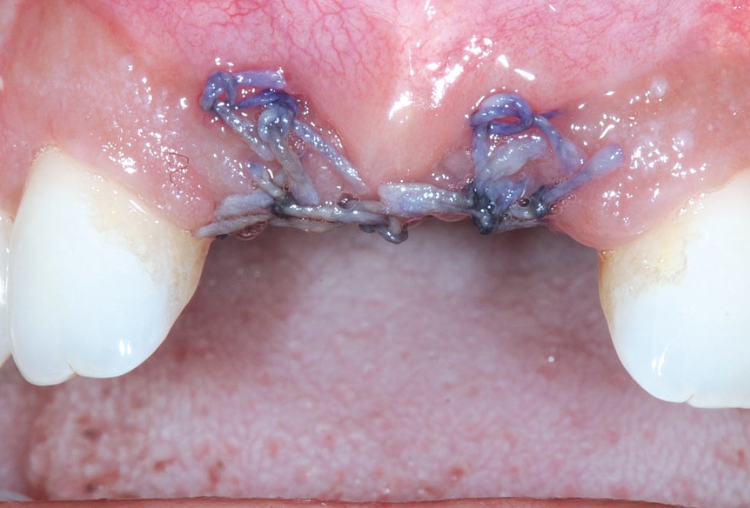
20/26 - 6 weeks post-op: sutures were removedBone augmentation in aesthetic zone with maxgraft® bonering - Dr. A. Patel
-
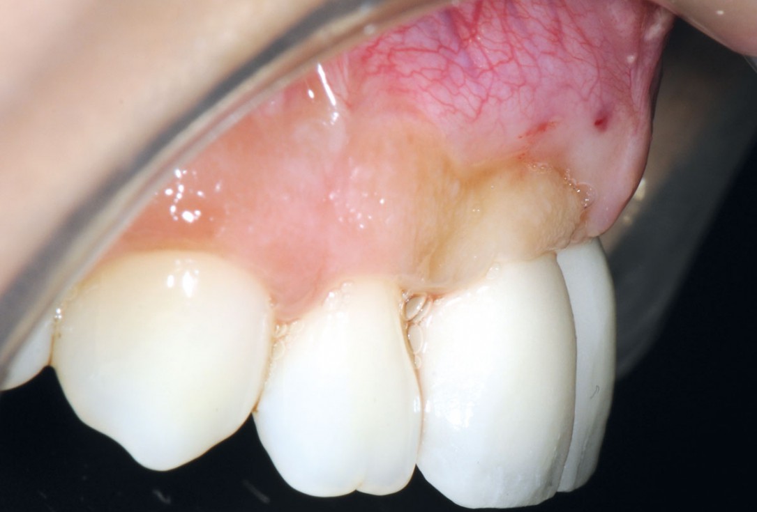
21/26 - 6 months after surgery: healthy soft tissuesBone augmentation in aesthetic zone with maxgraft® bonering - Dr. A. Patel
-
22/26 - Uncovering the implants 6 months after surgeryBone augmentation in aesthetic zone with maxgraft® bonering - Dr. A. Patel
-
23/26 - Prosthetic rehabilitationBone augmentation in aesthetic zone with maxgraft® bonering - Dr. A. Patel
-
24/26 - CT scan after implants been restoredBone augmentation in aesthetic zone with maxgraft® bonering - Dr. A. Patel
-
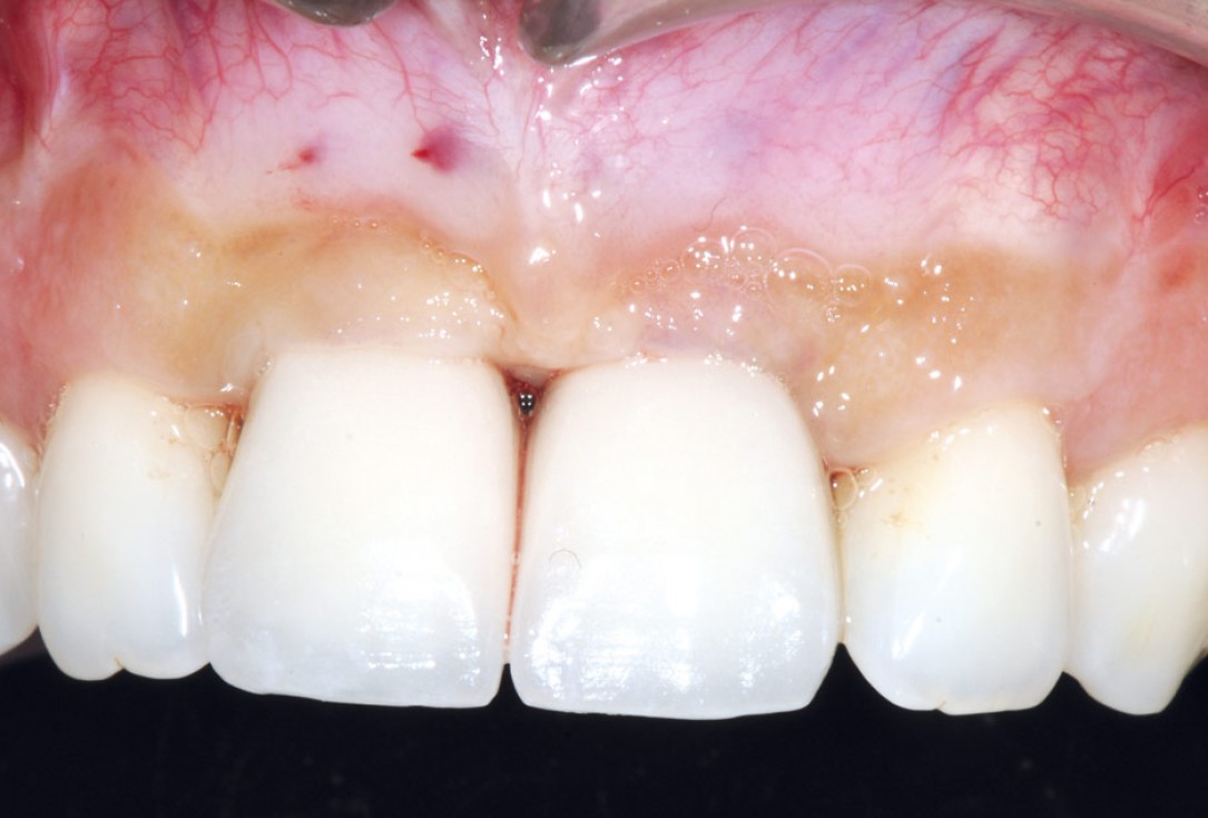
25/26 - Final crowns immediatly after restorationBone augmentation in aesthetic zone with maxgraft® bonering - Dr. A. Patel
-
26/26 - Lateral view confirms bone volume gainBone augmentation in aesthetic zone with maxgraft® bonering - Dr. A. Patel

Vertical augmentation: Preparation of ring bed in atrophic mandibula (third quadrant)

Initial situation: missing incisor with loss of buccal wall

Planning the surgery with CoDiagnostix® for Straumann® Guided Surgery

X-ray scan reveals initial situation with maxillary bone height in regio 15 of 1.5 mm

Initial situation: bone loss due to lack of physical load of bridge retained region 11

Initial situation pre-op: Central incisors with mobility 3

Severe periimplantitis at tooth 15 with bone loss up to 1/3 of the implant

Initial situation: X-ray scan reveals eggshell thin sinus floor (1-3 mm) on both sites of the maxilla; green areas indicate the planned maxgraft® bonerings and red areas the planned implants

Initially bridge retained incisors

X-ray scan of clinical situation

Initial situation 57-year old female patient. X-ray scan reveals severe bone loss due to inflammation in region 13. Treatment plan was extraction of teeth 13 and 14 and augmentation after healing.

X-ray scan: initial situation loss of two wall bony defect with loss of buccal and lingual lamella

Initial situation: single tooth gap in regio 22

Initial presentation of failing post retained crown with previous history of failed apicectomies and amalgam tattooing and scar tissue
