GBR with maxresorb® & Jason® membrane - Prof. Dr. Dr. D. Rothamel
-
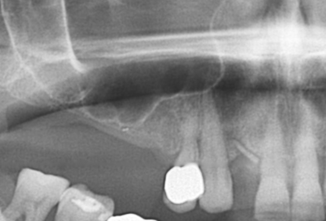
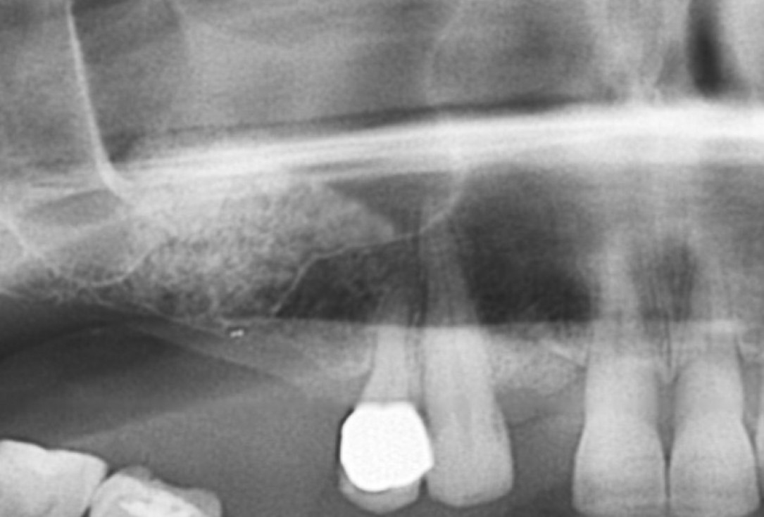
01/20 - Pre-operative x-rayGBR with maxresorb® & Jason® membrane - Prof. Dr. Dr. D. Rothamel
-
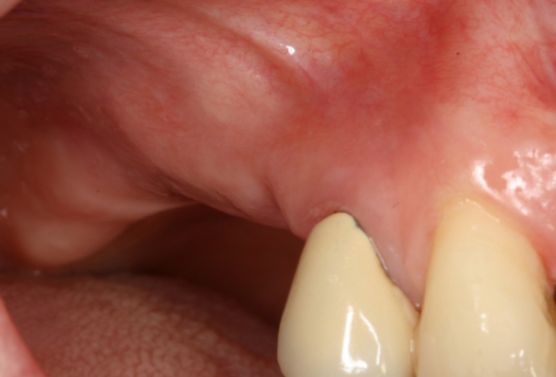
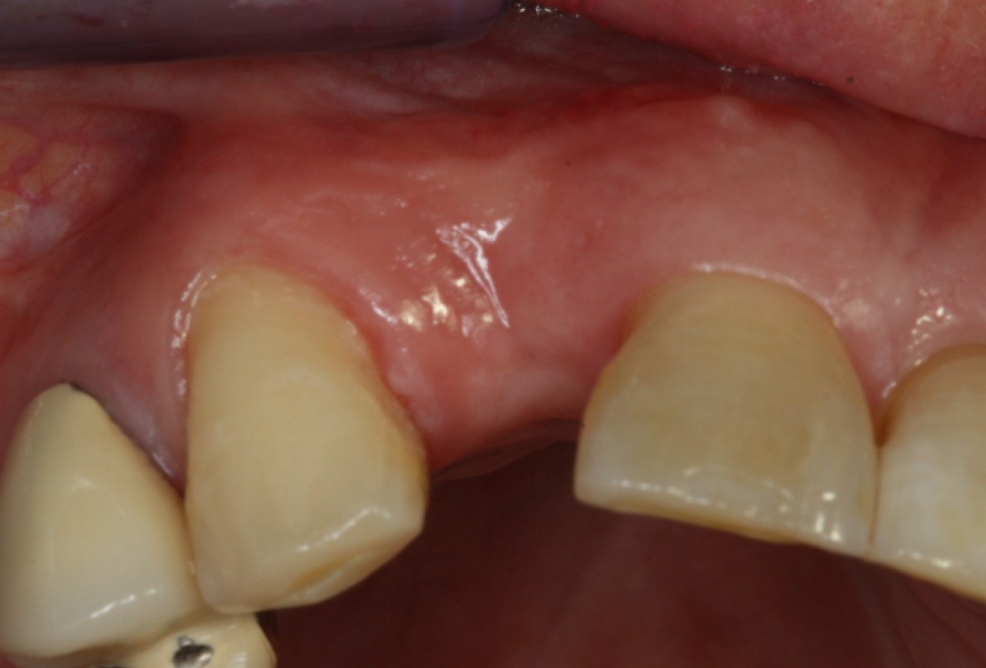
02/20 - Clinical situation before surgeryGBR with maxresorb® & Jason® membrane - Prof. Dr. Dr. D. Rothamel
-
03/20 - Lateral sinus window prepared and Schneiderian membrane protected with a Jason® membraneGBR with maxresorb® & Jason® membrane - Prof. Dr. Dr. D. Rothamel
-
04/20 - Filling of sinus cavity with maxresorb®GBR with maxresorb® & Jason® membrane - Prof. Dr. Dr. D. Rothamel
-
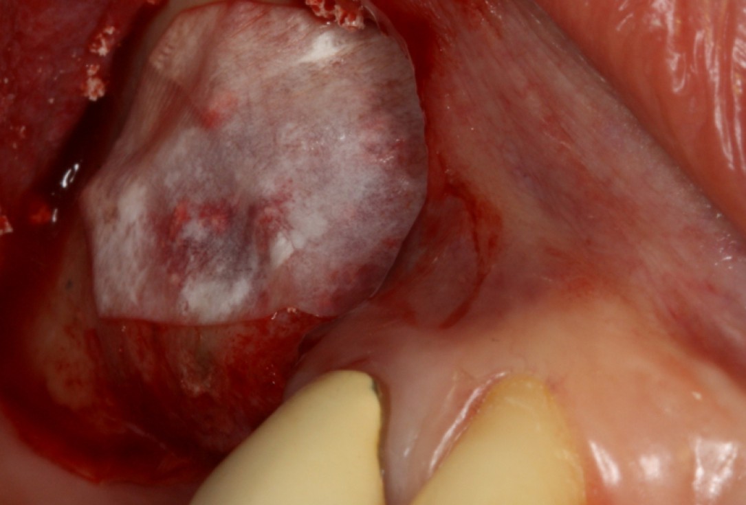
05/20 - Covering of augmentation site with the Jason® membraneGBR with maxresorb® & Jason® membrane - Prof. Dr. Dr. D. Rothamel
-
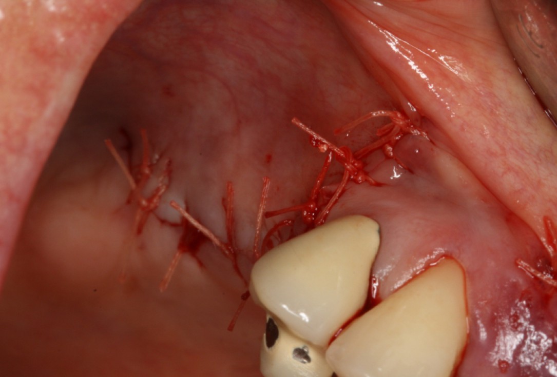
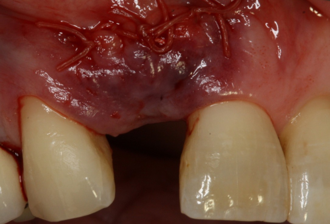
06/20 - Tension-free wound closureGBR with maxresorb® & Jason® membrane - Prof. Dr. Dr. D. Rothamel
-
07/20 - Second surgical site: clinical situation preoperativelyGBR with maxresorb® & Jason® membrane - Prof. Dr. Dr. D. Rothamel
-
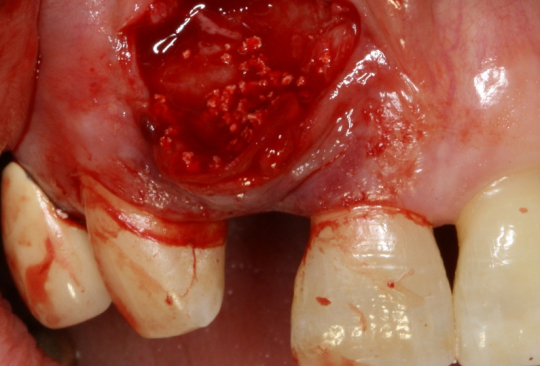
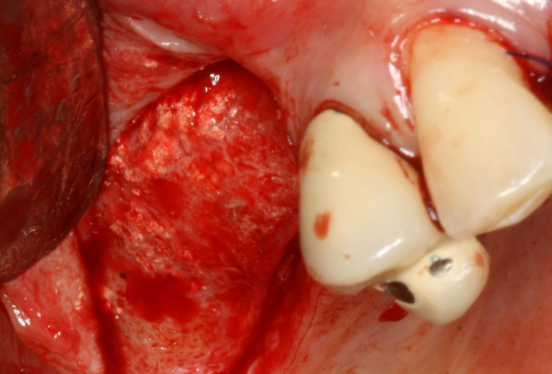
08/20 - Surgical presentation of the alveolar ridgeGBR with maxresorb® & Jason® membrane - Prof. Dr. Dr. D. Rothamel
-
09/20 - maxresorb® inserted into the extraction socketGBR with maxresorb® & Jason® membrane - Prof. Dr. Dr. D. Rothamel
-
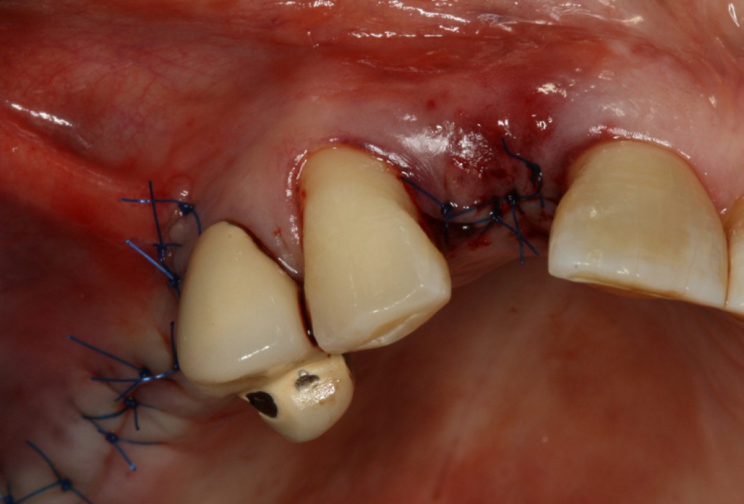
10/20 - wound closureGBR with maxresorb® & Jason® membrane - Prof. Dr. Dr. D. Rothamel
-
11/20 - Detail of OPG showing radiopacity of maxresorb®GBR with maxresorb® & Jason® membrane - Prof. Dr. Dr. D. Rothamel
-
12/20 - Good integration of maxresorb® particles at re-entryGBR with maxresorb® & Jason® membrane - Prof. Dr. Dr. D. Rothamel
-
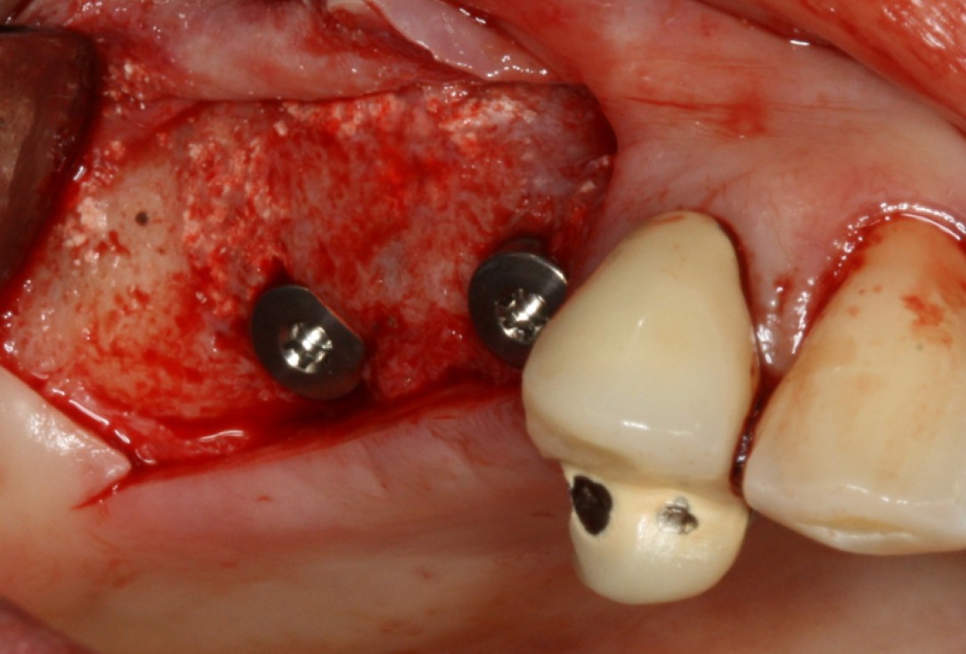
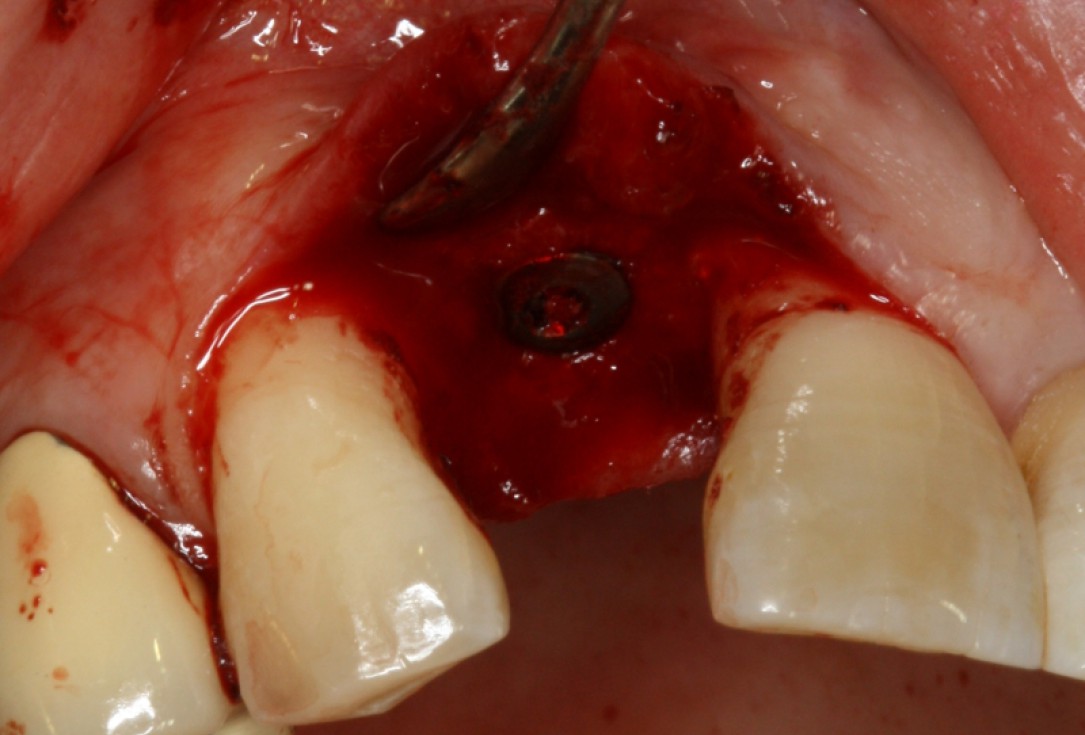
13/20 - Implant placementGBR with maxresorb® & Jason® membrane - Prof. Dr. Dr. D. Rothamel
-
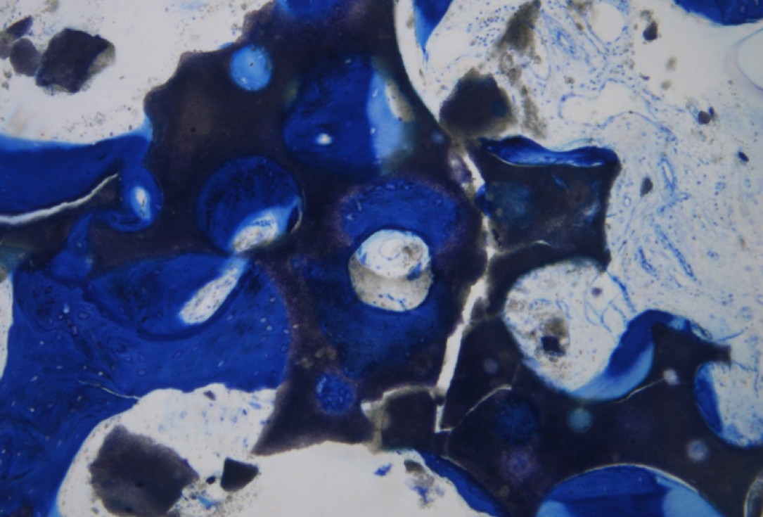
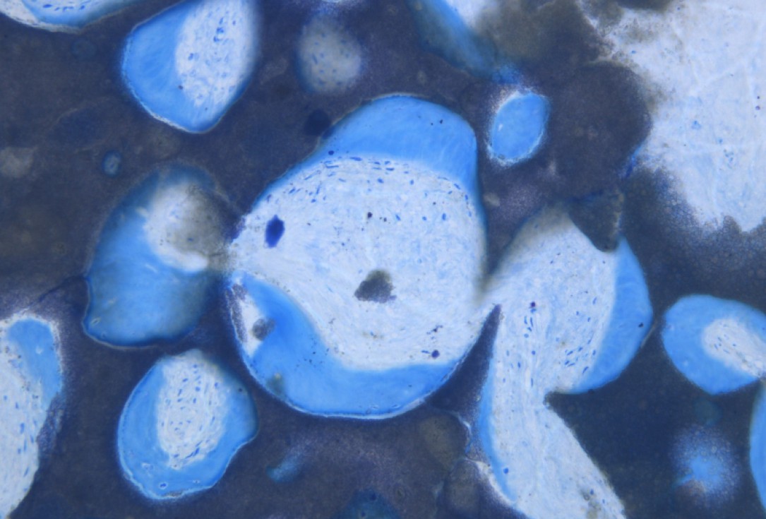
14/20 - Histology showing maxresorb® particles integrated in newly formed bone matrixGBR with maxresorb® & Jason® membrane - Prof. Dr. Dr. D. Rothamel
-
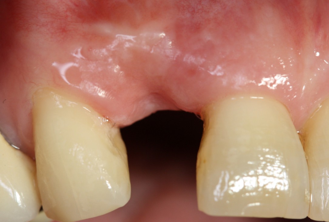
15/20 - Good soft tissue situation after healingGBR with maxresorb® & Jason® membrane - Prof. Dr. Dr. D. Rothamel
-
16/20 - re-entry: maxresorb® particles integrated into newly formed boneGBR with maxresorb® & Jason® membrane - Prof. Dr. Dr. D. Rothamel
-
17/20 - implant placementGBR with maxresorb® & Jason® membrane - Prof. Dr. Dr. D. Rothamel
-
18/20 - Histology showing the integration of maxresorb®GBR with maxresorb® & Jason® membrane - Prof. Dr. Dr. D. Rothamel
-
19/20 - SuturingGBR with maxresorb® & Jason® membrane - Prof. Dr. Dr. D. Rothamel
-
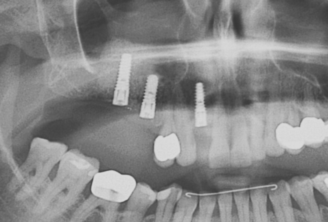
20/20 - OPG showing stable placement of implantsGBR with maxresorb® & Jason® membrane - Prof. Dr. Dr. D. Rothamel

Initial Orthopantomograph X-Ray

Pre-operative x-ray

X-ray control before tooth extraction

DVT image demonstrating horizontal and vertical amount of bone available

Surgical presentation of the alveolar ridge with reduced amount of horizontal bone available

DVT control after sinusitis surgery, residual bone height 1 mm

Clinical situation before extraction

DVT control after sinusitis surgery, residual bone height 1 mm

DVT image showing the reduced amount of bone available in the area of the mental foramen

X-ray shows a 3-dimensional periondontal defect

Initial situation: Inflammated tooth #12
