Soft tissue augmentation with mucoderm® for pontic - Dr. S. Stavar
-
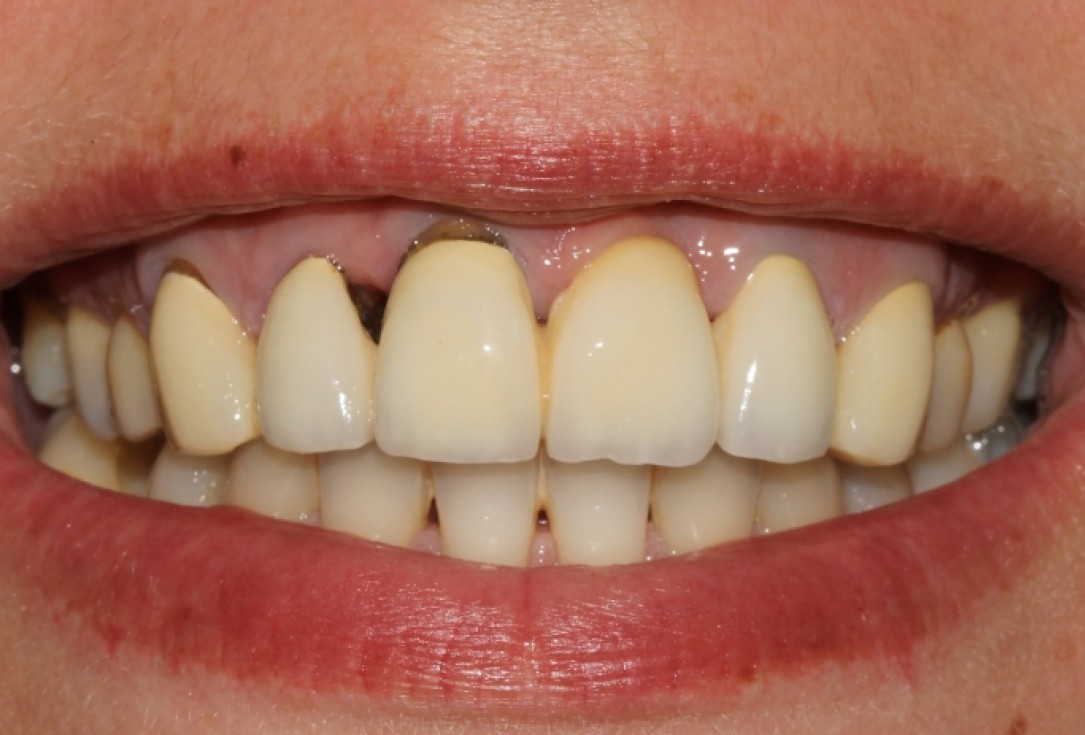
1/8 - Initial clinical situationSoft tissue augmentation with mucoderm® for pontic - Dr. S. Stavar
-
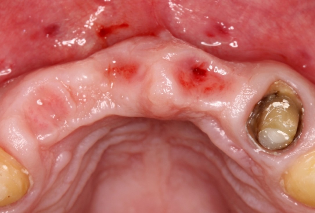
2/8 - Clinical view after removal of the temporary bridgeSoft tissue augmentation with mucoderm® for pontic - Dr. S. Stavar
-
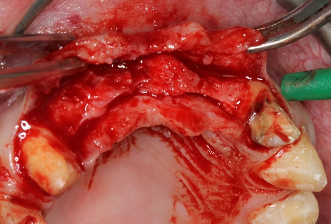
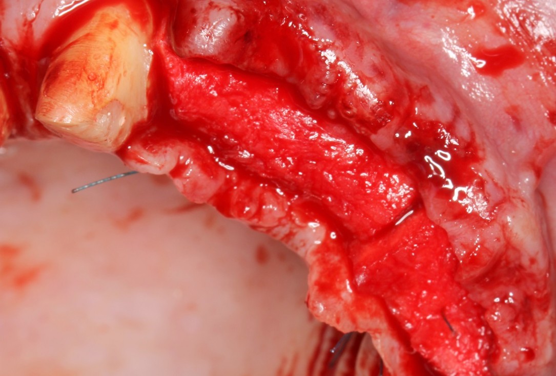
3/8 - Situation after mobilization of a mucosal flapSoft tissue augmentation with mucoderm® for pontic - Dr. S. Stavar
-
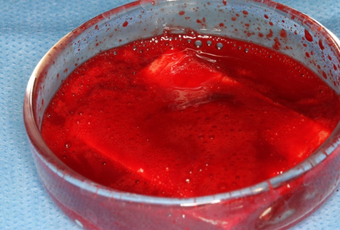
4/8 - Rehydration of mucoderm® in bloodSoft tissue augmentation with mucoderm® for pontic - Dr. S. Stavar
-
5/8 - mucoderm® in situ fixed by suturesSoft tissue augmentation with mucoderm® for pontic - Dr. S. Stavar
-
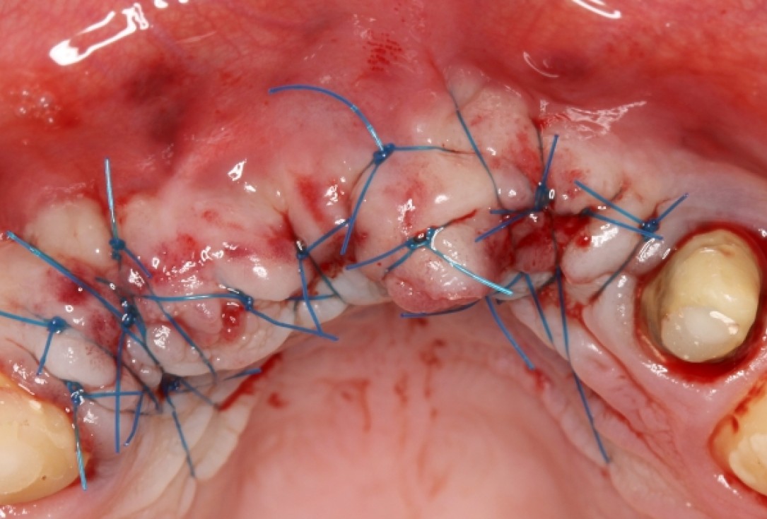
6/8 - Wound closure and suturingSoft tissue augmentation with mucoderm® for pontic - Dr. S. Stavar
-
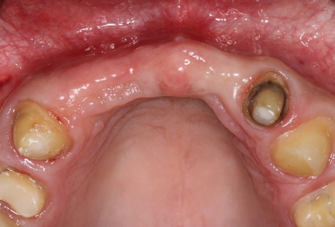
7/8 - Clinical situation 3 months post-operativeSoft tissue augmentation with mucoderm® for pontic - Dr. S. Stavar
-
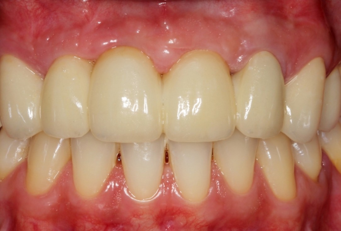
8/8 - Final prosthetic restorationSoft tissue augmentation with mucoderm® for pontic - Dr. S. Stavar

X-ray of initial clinical situation

Initial clinical situation showing severe soft tissue loss

Initial clinical situation

Full-thickness flap preparation bucally and lingually

Initial clinical situation

Initial clinical situation

Initial clinical situation with narrow ridge

Initial clinical situation showing tooth 45 not worth preserving

Drilling template for guided implant placement

recession on tooth 11

Initial clinical situation

X-ray shows a 3-dimensional periondontal defect

Bone defect in area 11-21 due to two lost implants (periimplantitis) after 15 years of function

Probing demonstrates peri-implant pocket depth of 8 mm
