cerabone® and mucoderm® for immediate implantation in the aesthetic area - Dr. D. Robles
-
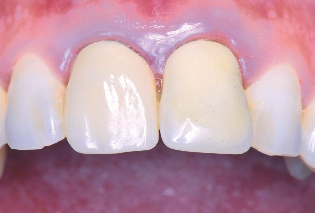
01/22 - Initial clinical situation - Central incisors with dental destruction and periapical pathologycerabone® and mucoderm® for immediate implantation in the aesthetic area - Dr. D. Robles
-
02/22 - Initial clinical situation - Central incisors with dental destruction and periapical pathologycerabone® and mucoderm® for immediate implantation in the aesthetic area - Dr. D. Robles
-
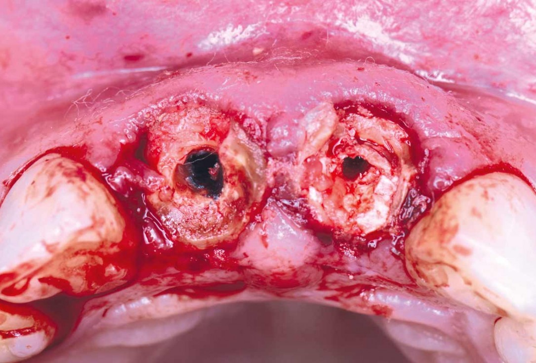
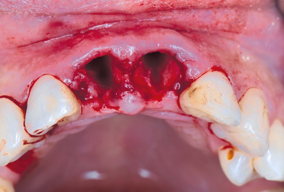
03/22 - Initial clinical situation – Tooth extractioncerabone® and mucoderm® for immediate implantation in the aesthetic area - Dr. D. Robles
-
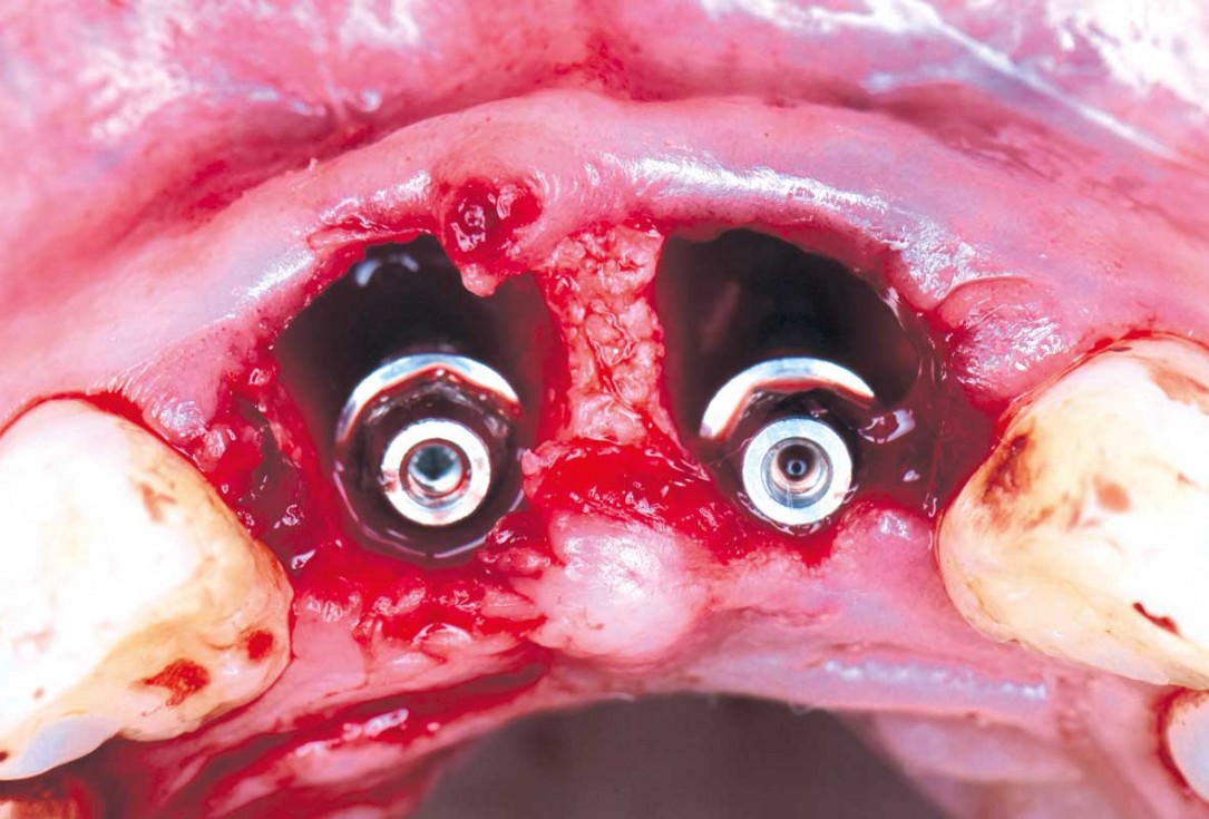
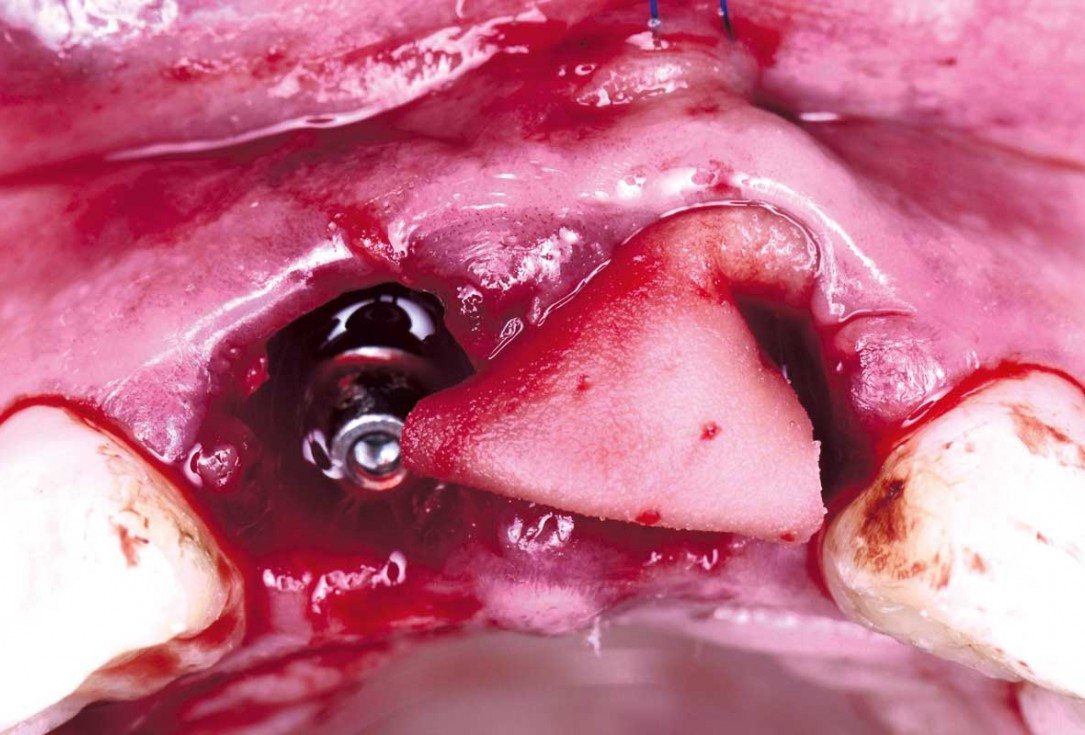
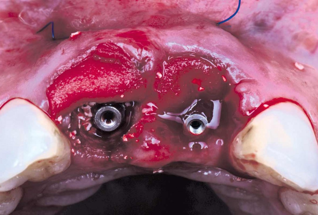
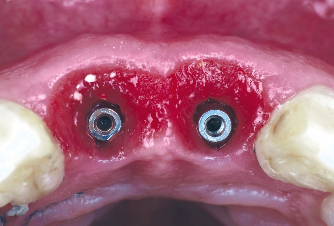
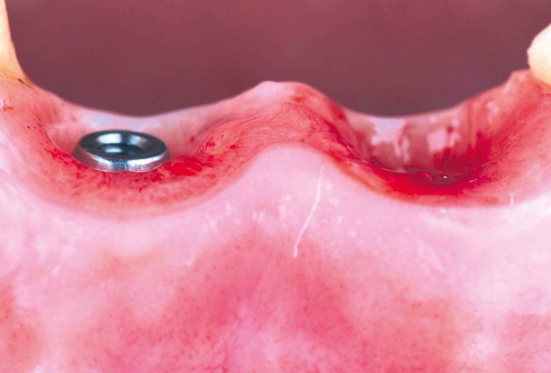
04/22 - Immediate implants are placed after extractioncerabone® and mucoderm® for immediate implantation in the aesthetic area - Dr. D. Robles
-
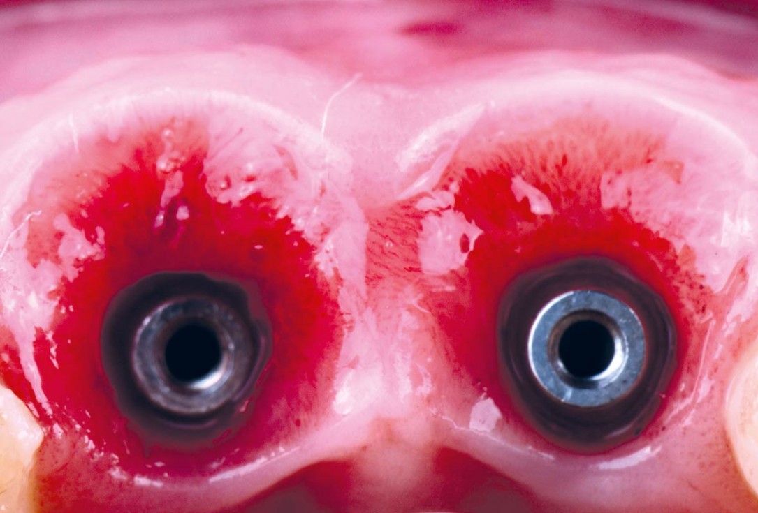
05/22 - Immediate implants are placed after extraction and the gap is filled with cerabone®cerabone® and mucoderm® for immediate implantation in the aesthetic area - Dr. D. Robles
-
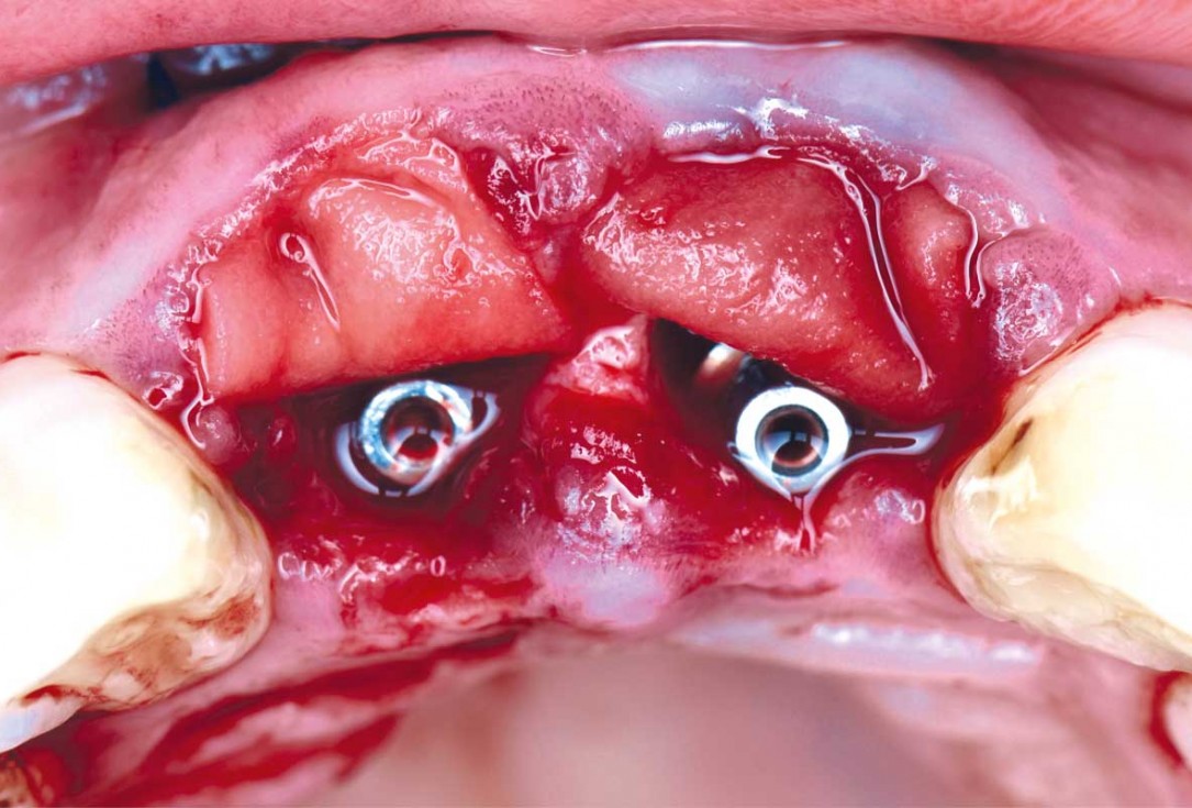
06/22 - To improve the soft tissues, two folded mucoderm® are placedcerabone® and mucoderm® for immediate implantation in the aesthetic area - Dr. D. Robles
-
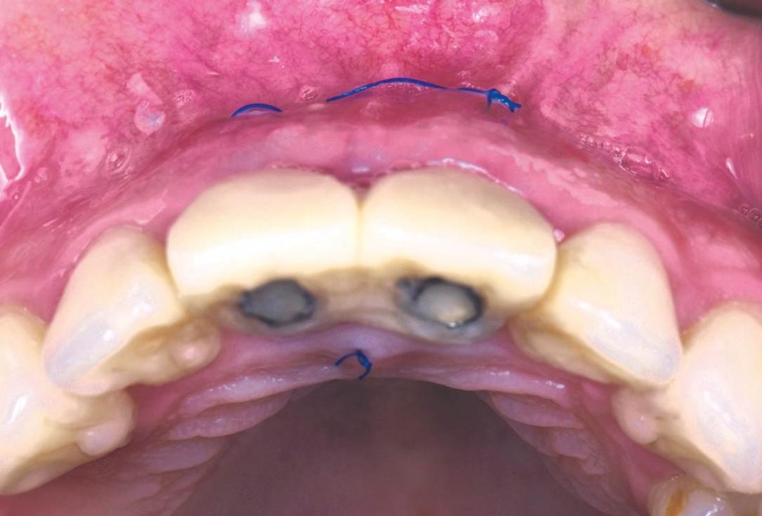
07/22 - To achieve greater volume, mucoderm® are placed through an envelope and sutured apicallycerabone® and mucoderm® for immediate implantation in the aesthetic area - Dr. D. Robles
-
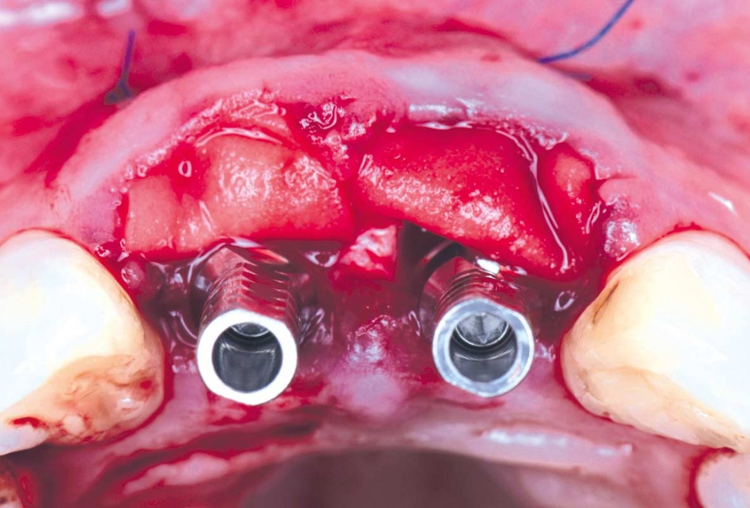
08/22 - To achieve greater volume, mucoderm® are placed through an envelope and sutured apicallycerabone® and mucoderm® for immediate implantation in the aesthetic area - Dr. D. Robles
-
09/22 - Gap is filled with cerabone®cerabone® and mucoderm® for immediate implantation in the aesthetic area - Dr. D. Robles
-
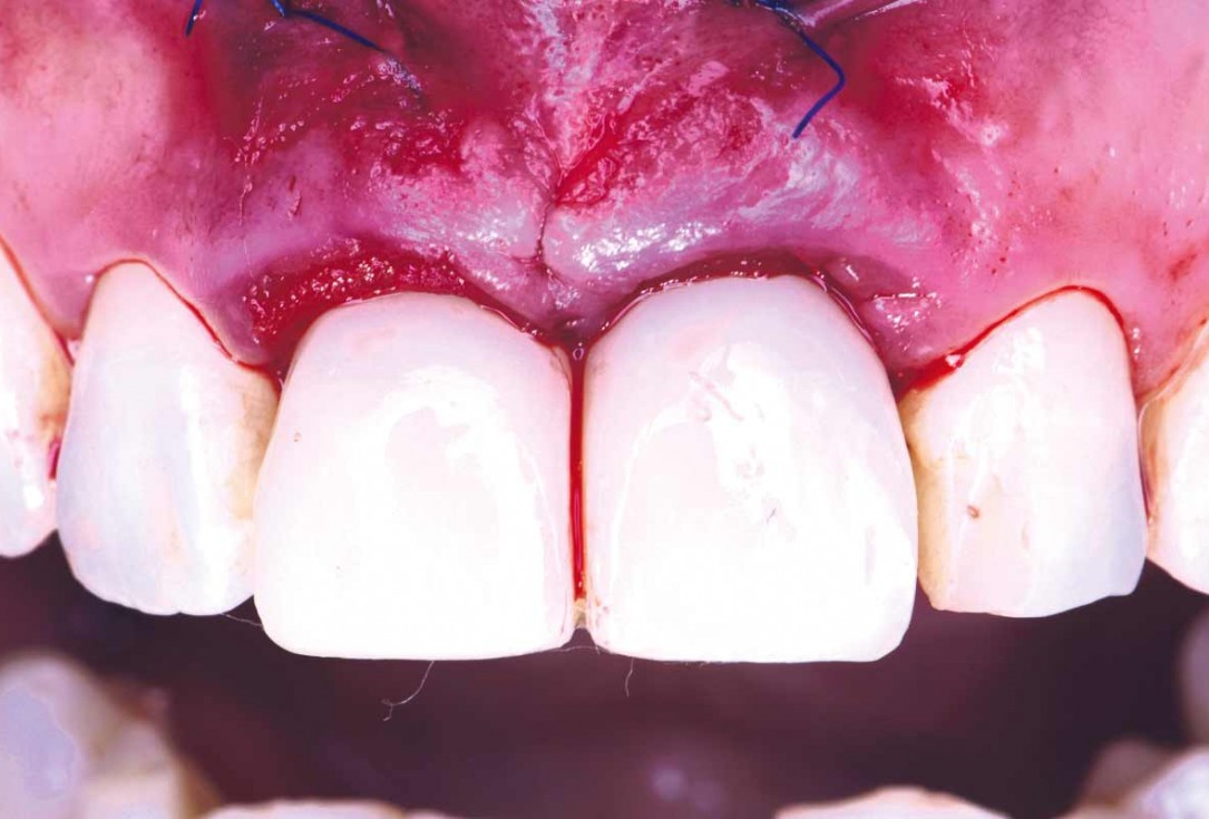
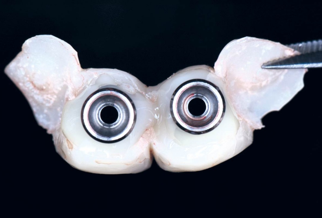
10/22 - Immediate provisional screw-retained prosthesis is placed to stabilize both biomaterials in the coronal areacerabone® and mucoderm® for immediate implantation in the aesthetic area - Dr. D. Robles
-
11/22 - Immediate provisional screw-retained prosthesiscerabone® and mucoderm® for immediate implantation in the aesthetic area - Dr. D. Robles
-
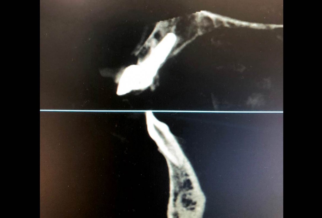
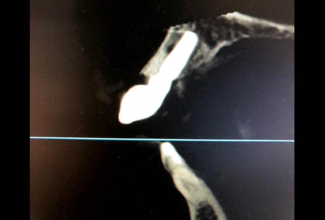
12/22 - Control X-Raycerabone® and mucoderm® for immediate implantation in the aesthetic area - Dr. D. Robles
-
13/22 - Control X-Raycerabone® and mucoderm® for immediate implantation in the aesthetic area - Dr. D. Robles
-
14/22 - Control X-Raycerabone® and mucoderm® for immediate implantation in the aesthetic area - Dr. D. Robles
-
15/22 - Follow up after 15 dayscerabone® and mucoderm® for immediate implantation in the aesthetic area - Dr. D. Robles
-
16/22 - Follow up after 15 days – Uneventful healing of the gumcerabone® and mucoderm® for immediate implantation in the aesthetic area - Dr. D. Robles
-
17/22 - Follow up after 5 months – Soft tissue maturationcerabone® and mucoderm® for immediate implantation in the aesthetic area - Dr. D. Robles
-
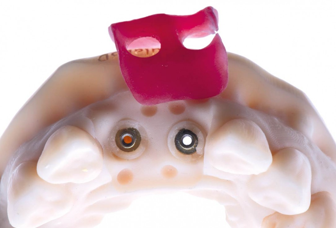
18/22 - CAD/CAM designed prosthesis after digital scan of tissuescerabone® and mucoderm® for immediate implantation in the aesthetic area - Dr. D. Robles
-
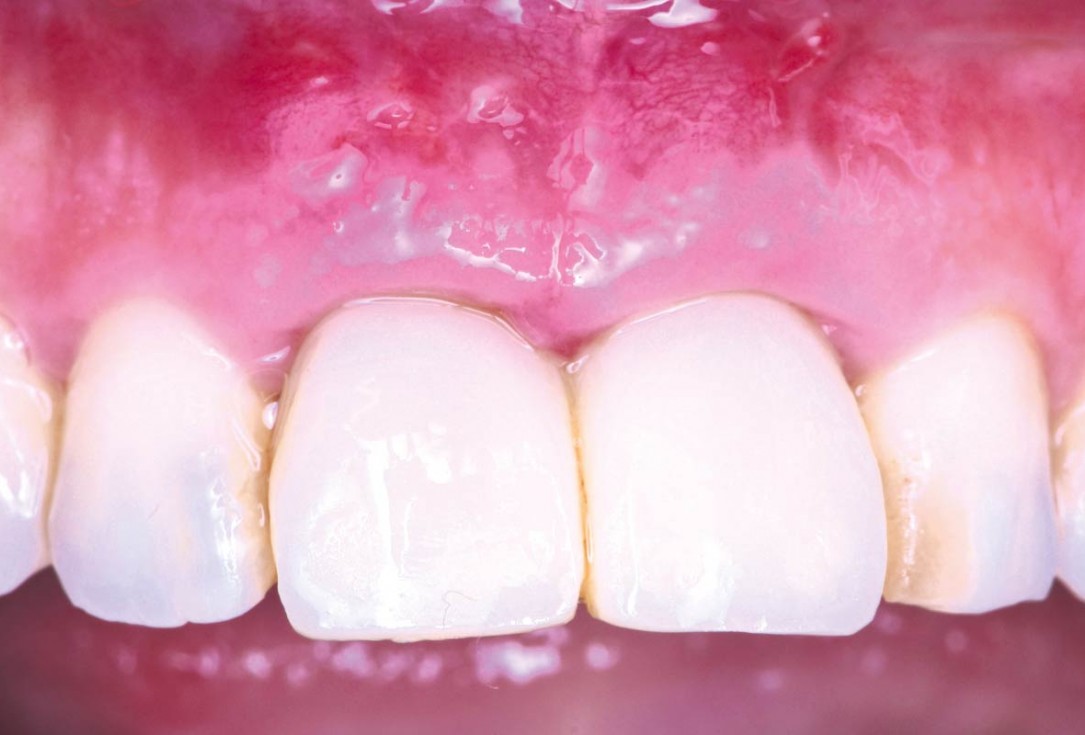
19/22 - Final clinical situation: 10 months follow up - aesthetic rehabilitationcerabone® and mucoderm® for immediate implantation in the aesthetic area - Dr. D. Robles
-
20/22 - Final clinical situation - aesthetic rehabilitationcerabone® and mucoderm® for immediate implantation in the aesthetic area - Dr. D. Robles
-
21/22 - Final clinical situation - aesthetic rehabilitationcerabone® and mucoderm® for immediate implantation in the aesthetic area - Dr. D. Robles
-
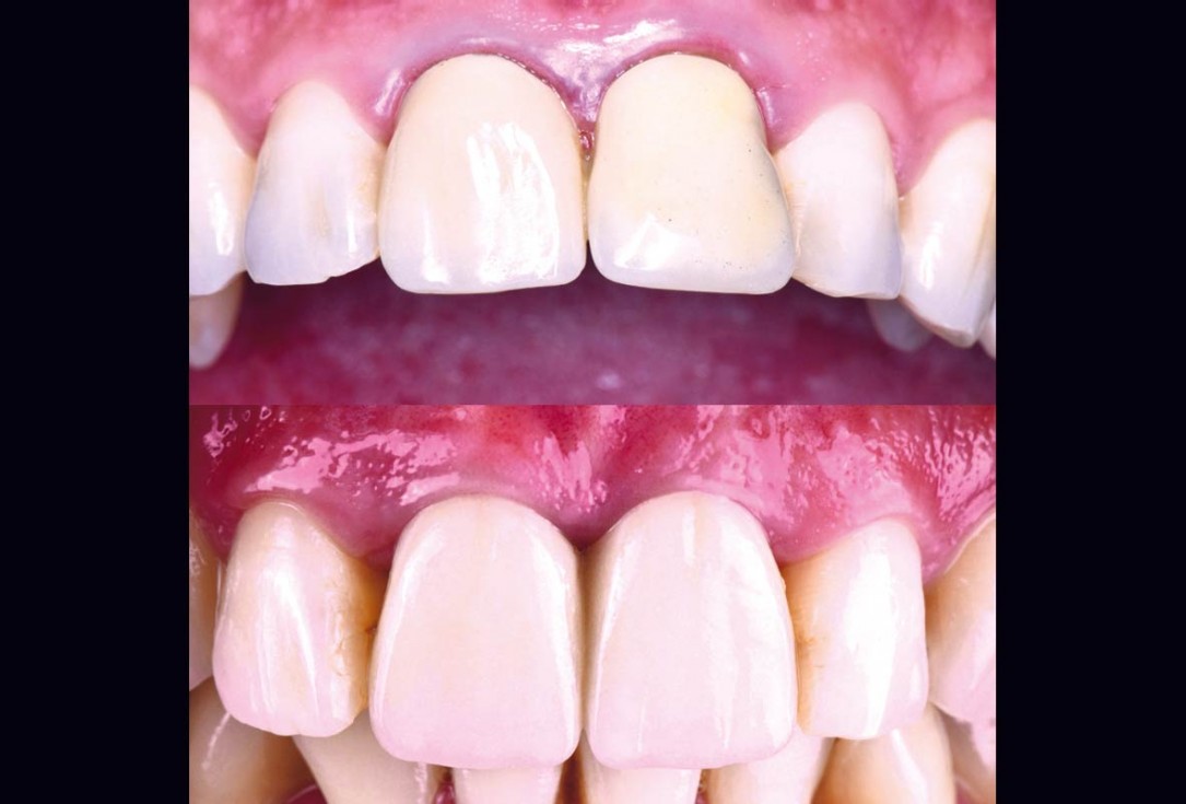
22/22 - Comparison before and after rehabilitationcerabone® and mucoderm® for immediate implantation in the aesthetic area - Dr. D. Robles

Initial Orthopantomograph X-Ray

Clinical situation before extraction and implantation

X-ray control before tooth extraction

Clinical view of the case.

Pre-operative x-ray image, teeth 43, 44, 45, 46 and 47 planned for extraction

Initial situation with broken tooth 46

47 years old patient referred by another dentist after suffering a fall while fishing

Preoperative Ortopantomogram of the teeth planned for extraction

Initial situation pre-op: Central incisors with mobility 3

Pre-operative OPG, tooth 36 planned for extraction

Initial situation with fractured central incisors

The patient presented with a terminal fracture of the crown tooth number 12

Immediately placed implant covered with permamem®. permamem® passively immobilized by sutures and intentionally left exposed to the oral cavity.

Pre-operative situation showing tooth 21 with deep periodontal pocket. Tooth presented with mobility grade III.

Initial view of the case. Discoloration of 1.1 and mild class I gingival recession

Pre-operative OPG, teeth 24, 25, and 26 planned for extraction
