GBR with maxresorb® & Jason® membrane - Prof. Dr. Dr. D. Rothamel
-
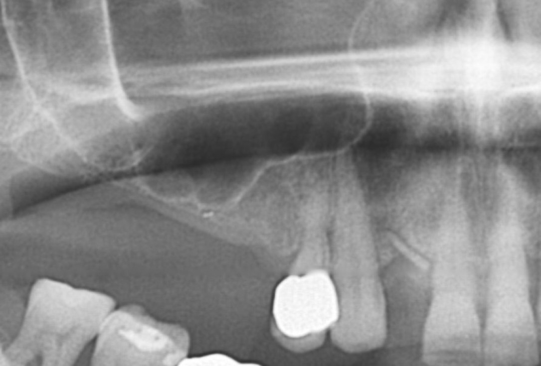
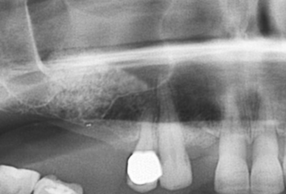
01/20 - Pre-operative x-rayGBR with maxresorb® & Jason® membrane - Prof. Dr. Dr. D. Rothamel
-
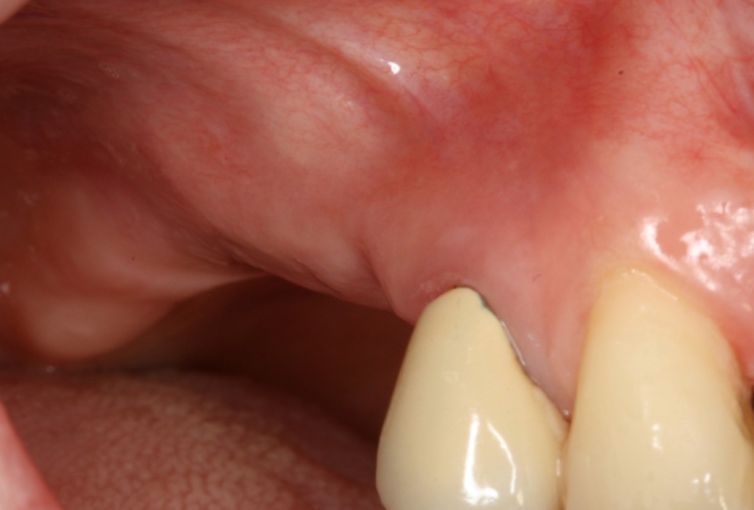
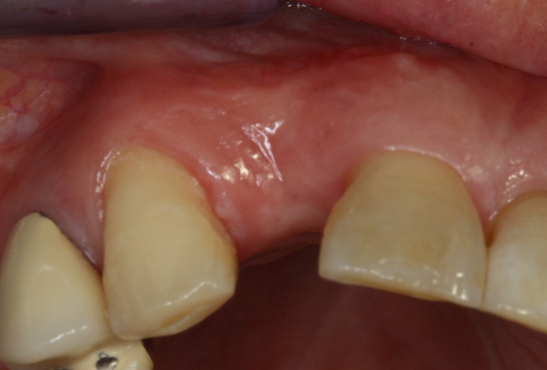
02/20 - Clinical situation before surgeryGBR with maxresorb® & Jason® membrane - Prof. Dr. Dr. D. Rothamel
-
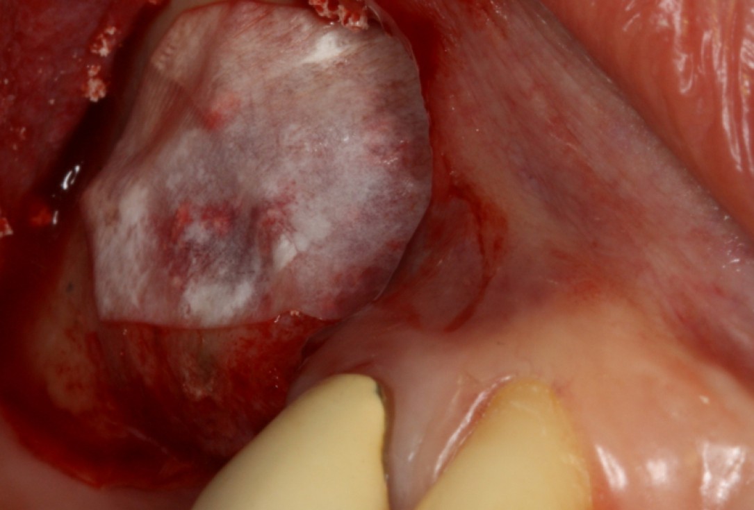
03/20 - Lateral sinus window prepared and Schneiderian membrane protected with a Jason® membraneGBR with maxresorb® & Jason® membrane - Prof. Dr. Dr. D. Rothamel
-
04/20 - Filling of sinus cavity with maxresorb®GBR with maxresorb® & Jason® membrane - Prof. Dr. Dr. D. Rothamel
-
05/20 - Covering of augmentation site with the Jason® membraneGBR with maxresorb® & Jason® membrane - Prof. Dr. Dr. D. Rothamel
-
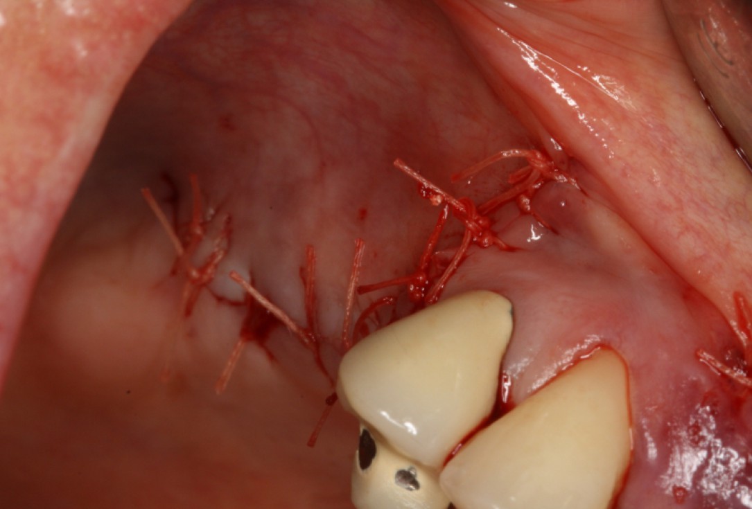
06/20 - Tension-free wound closureGBR with maxresorb® & Jason® membrane - Prof. Dr. Dr. D. Rothamel
-
07/20 - Second surgical site: clinical situation preoperativelyGBR with maxresorb® & Jason® membrane - Prof. Dr. Dr. D. Rothamel
-
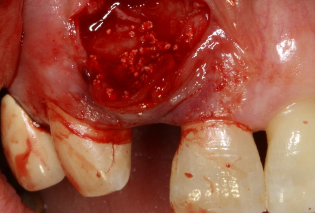
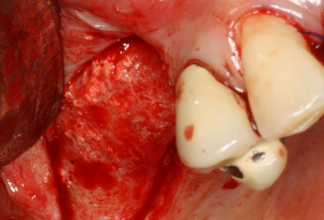
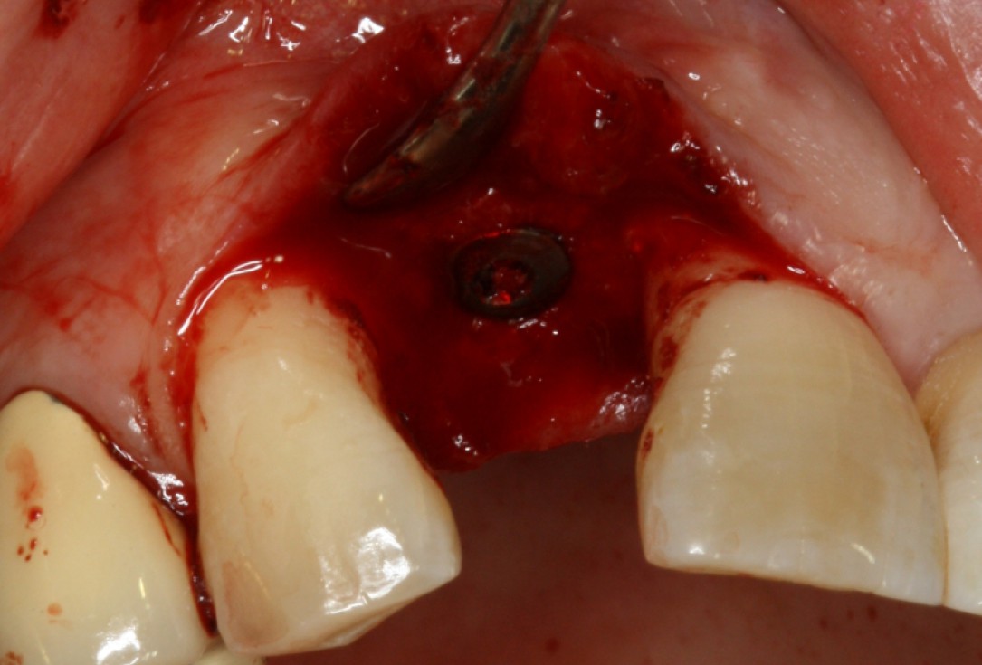
08/20 - Surgical presentation of the alveolar ridgeGBR with maxresorb® & Jason® membrane - Prof. Dr. Dr. D. Rothamel
-
09/20 - maxresorb® inserted into the extraction socketGBR with maxresorb® & Jason® membrane - Prof. Dr. Dr. D. Rothamel
-
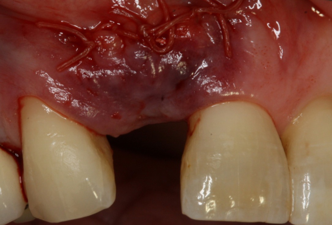
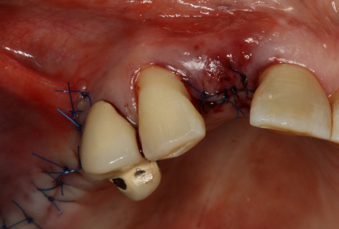
10/20 - wound closureGBR with maxresorb® & Jason® membrane - Prof. Dr. Dr. D. Rothamel
-
11/20 - Detail of OPG showing radiopacity of maxresorb®GBR with maxresorb® & Jason® membrane - Prof. Dr. Dr. D. Rothamel
-
12/20 - Good integration of maxresorb® particles at re-entryGBR with maxresorb® & Jason® membrane - Prof. Dr. Dr. D. Rothamel
-
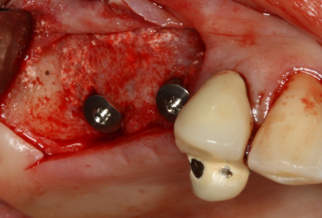
13/20 - Implant placementGBR with maxresorb® & Jason® membrane - Prof. Dr. Dr. D. Rothamel
-
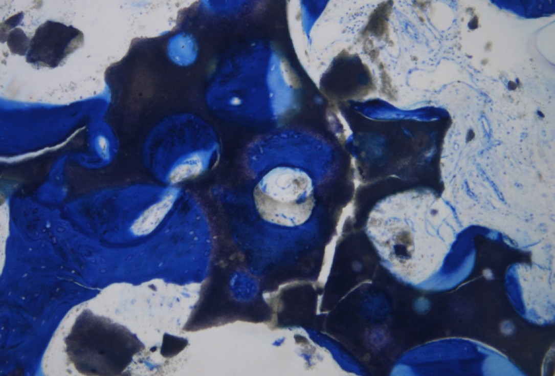
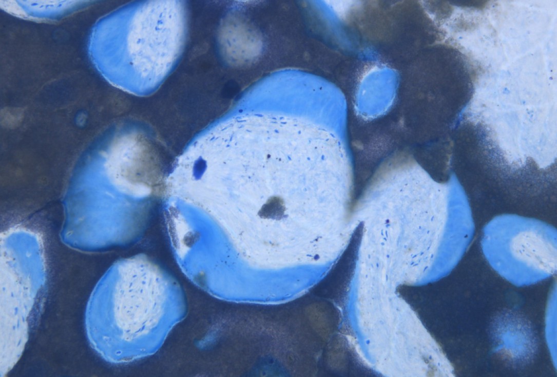
14/20 - Histology showing maxresorb® particles integrated in newly formed bone matrixGBR with maxresorb® & Jason® membrane - Prof. Dr. Dr. D. Rothamel
-
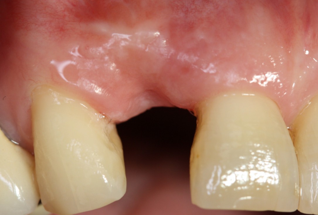
15/20 - Good soft tissue situation after healingGBR with maxresorb® & Jason® membrane - Prof. Dr. Dr. D. Rothamel
-
16/20 - re-entry: maxresorb® particles integrated into newly formed boneGBR with maxresorb® & Jason® membrane - Prof. Dr. Dr. D. Rothamel
-
17/20 - implant placementGBR with maxresorb® & Jason® membrane - Prof. Dr. Dr. D. Rothamel
-
18/20 - Histology showing the integration of maxresorb®GBR with maxresorb® & Jason® membrane - Prof. Dr. Dr. D. Rothamel
-
19/20 - SuturingGBR with maxresorb® & Jason® membrane - Prof. Dr. Dr. D. Rothamel
-
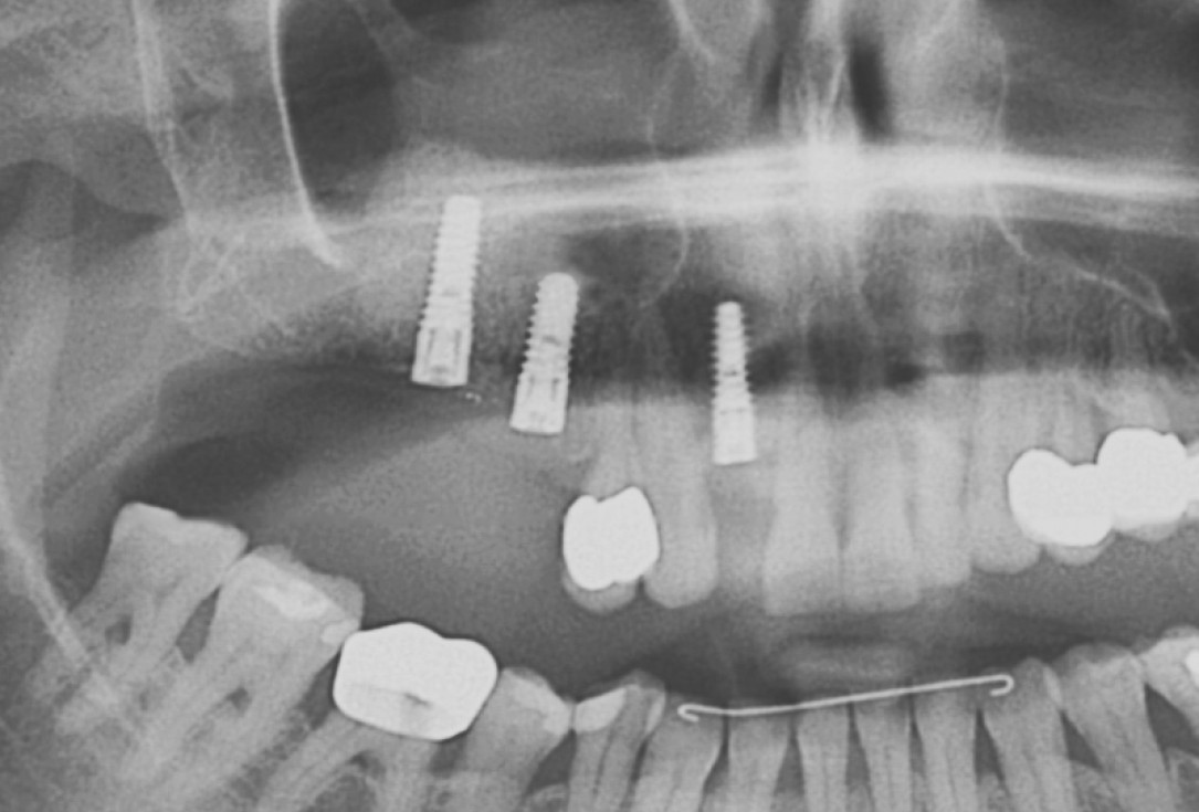
20/20 - OPG showing stable placement of implantsGBR with maxresorb® & Jason® membrane - Prof. Dr. Dr. D. Rothamel

Initial Orthopantomograph X-Ray

DVT image showing the reduced amount of bone available in the area of the mental foramen

X-ray control before tooth extraction

Initial situation: Inflammated tooth #12

Surgical presentation of the alveolar ridge with reduced amount of horizontal bone available

DVT control after sinusitis surgery, residual bone height 1 mm

Clinical situation before extraction

DVT image demonstrating horizontal and vertical amount of bone available

Surgical presentation of the alveolar ridge with reduced amount of horizontal bone available

X-ray shows a 3-dimensional periondontal defect

X-ray control showing initial situation

DVT control after sinusitis surgery, residual bone height 1 mm
