Bone augementation with maxresorb® - Dr. R. Cutts
-
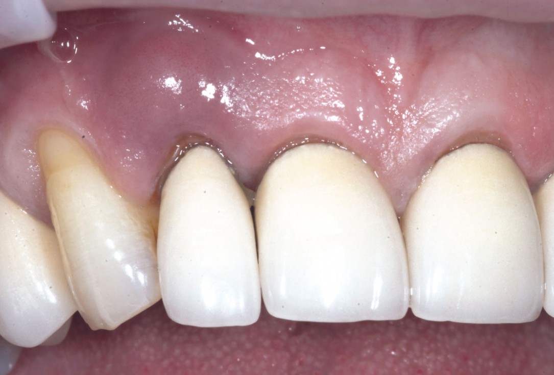
1/19 - Initial situation: Inflammated tooth #12Bone augementation with maxresorb® - Dr. R. Cutts
-
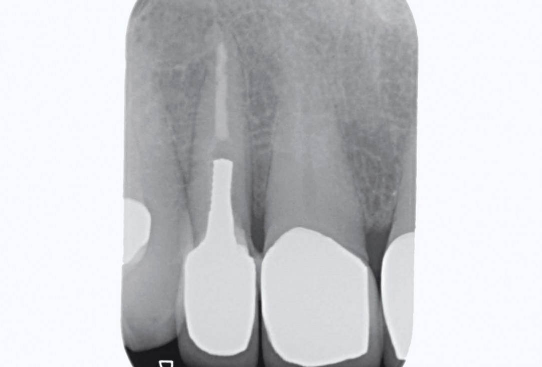
2/19 - Initial situation: x-rayBone augementation with maxresorb® - Dr. R. Cutts
-
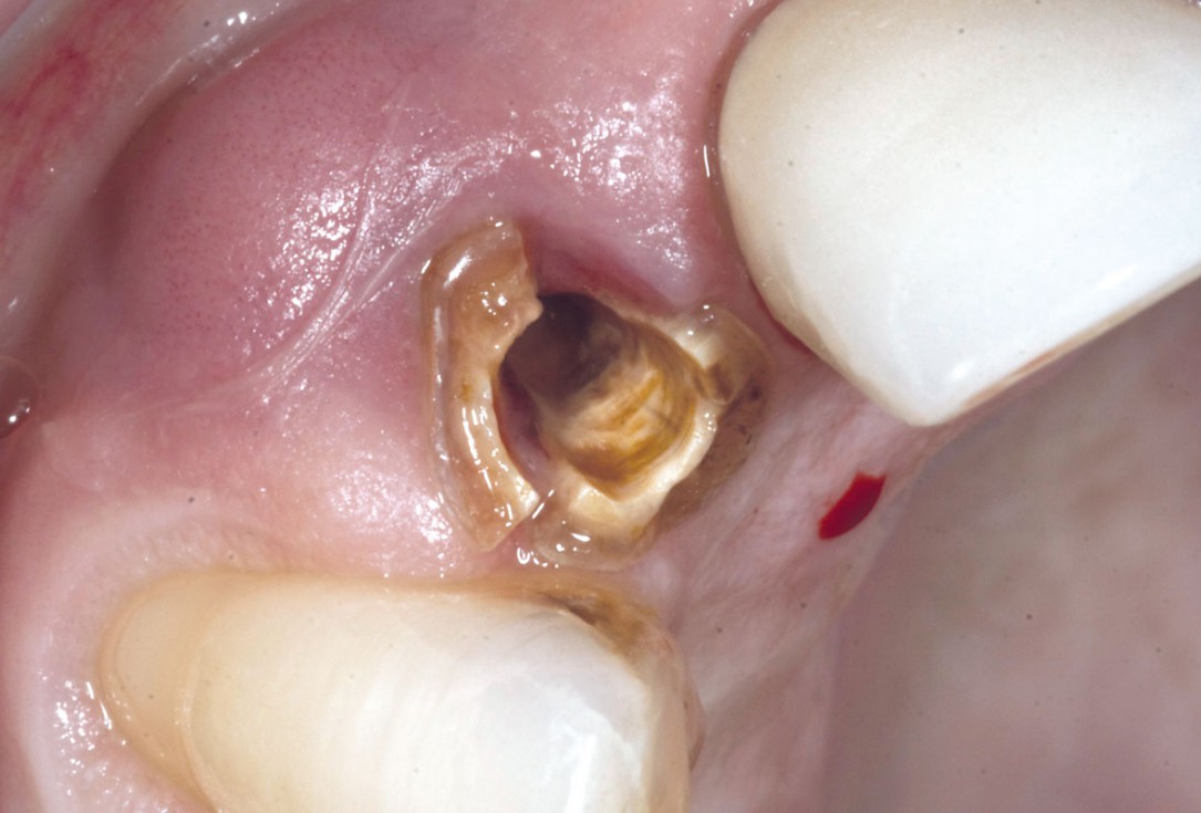
3/19 - Fracture of tooth while extractionBone augementation with maxresorb® - Dr. R. Cutts
-
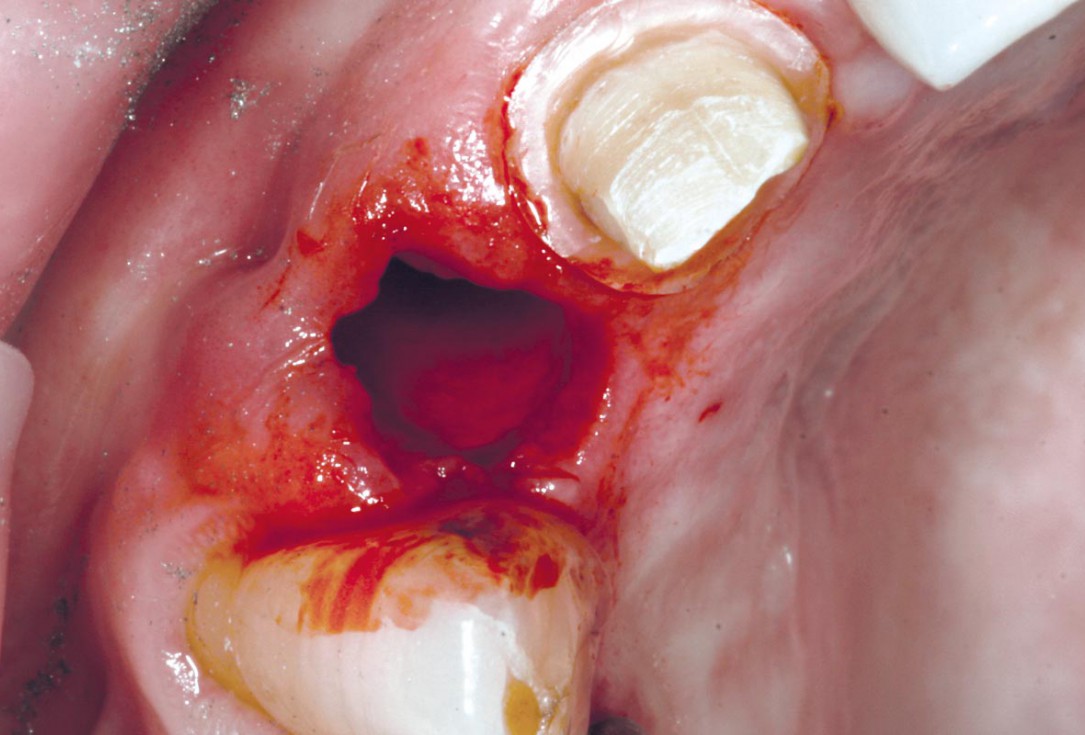
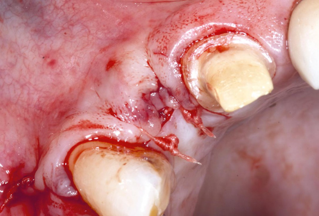
4/19 - Site after extractionBone augementation with maxresorb® - Dr. R. Cutts
-
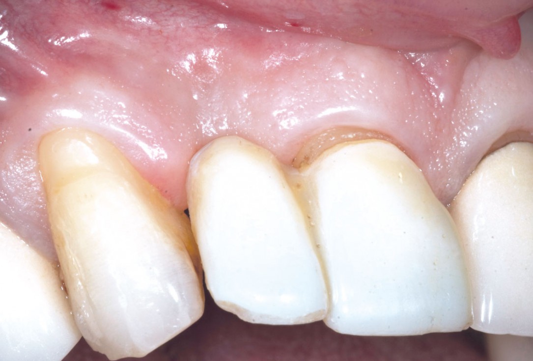
5/19 - 7 weeks after extraction of healing with a temporary tooth supported cantilever bridgeBone augementation with maxresorb® - Dr. R. Cutts
-
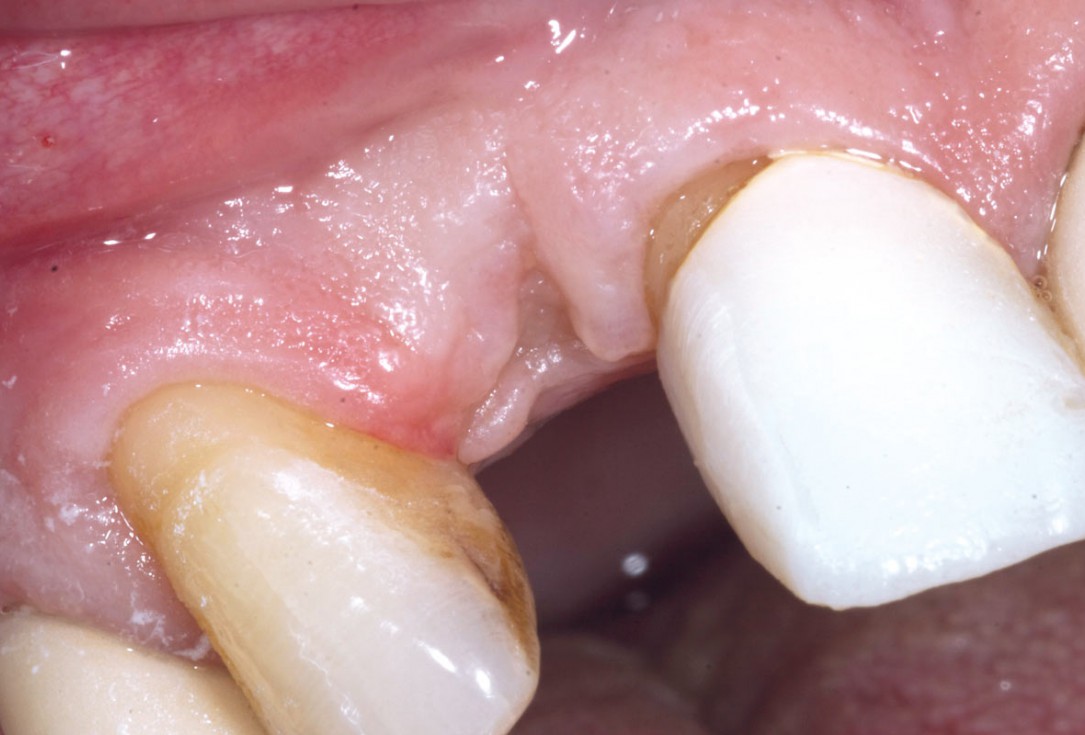
6/19 - Site without prosthetics showing sever buccal bone lossBone augementation with maxresorb® - Dr. R. Cutts
-
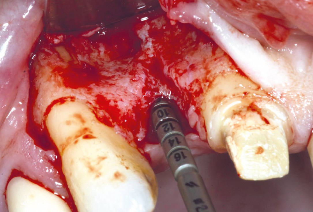
7/19 - Angulation and length of the dental implant measuredBone augementation with maxresorb® - Dr. R. Cutts
-
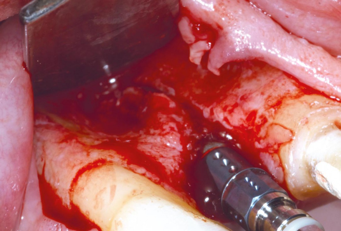
8/19 - Placement of Straumann BL implantBone augementation with maxresorb® - Dr. R. Cutts
-
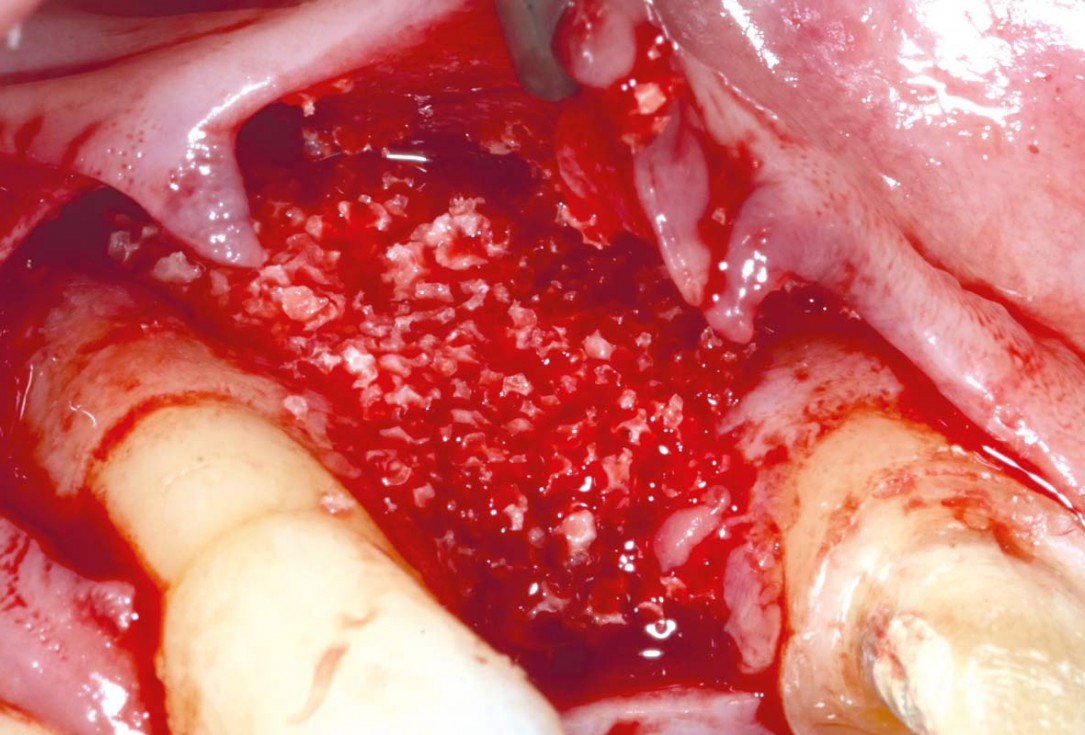
9/19 - Bone grafting with maxresorb® granulesBone augementation with maxresorb® - Dr. R. Cutts
-
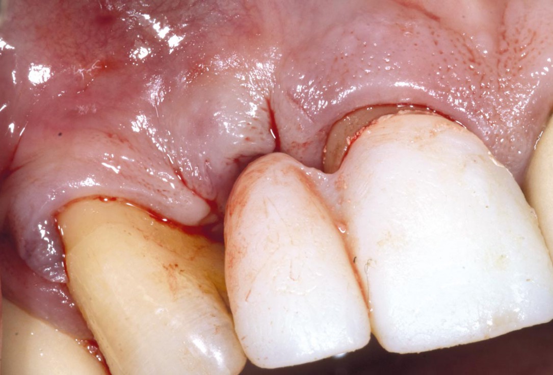
10/19 - Flap closed with single suturesBone augementation with maxresorb® - Dr. R. Cutts
-
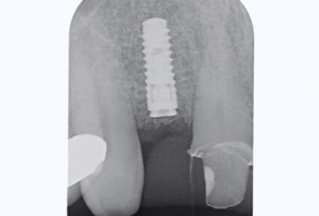
11/19 - Post-op x-ray shows good seating of implantBone augementation with maxresorb® - Dr. R. Cutts
-
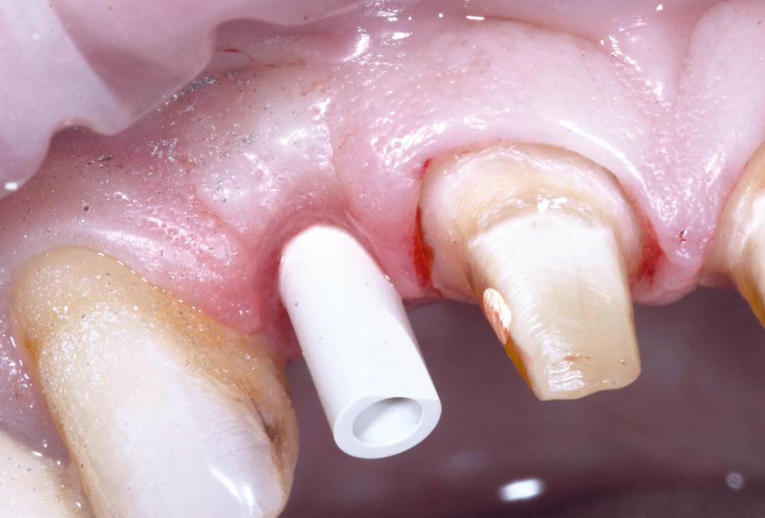
12/19 - Temporary restoration during healing phaseBone augementation with maxresorb® - Dr. R. Cutts
-
13/19 - 7 weeks after implant placementBone augementation with maxresorb® - Dr. R. Cutts
-
14/19 - Scanning the Implant fixture in situ with Digital Itero Scan including repreparation of adjacent teethBone augementation with maxresorb® - Dr. R. Cutts
-
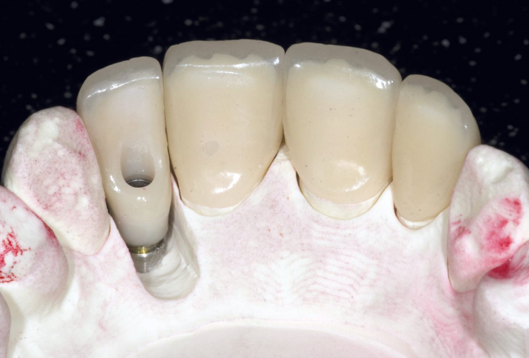
15/19 - Screw retained final implant prosthesis and adjacent ceramic unitsBone augementation with maxresorb® - Dr. R. Cutts
-
16/19 - Delivery of final prostheticsBone augementation with maxresorb® - Dr. R. Cutts
-
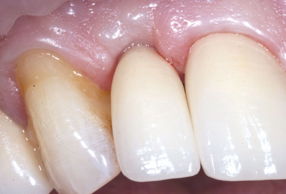
17/19 - 2 weeks after delivery of final prosthesis and 10 weeks after implant placementBone augementation with maxresorb® - Dr. R. Cutts
-
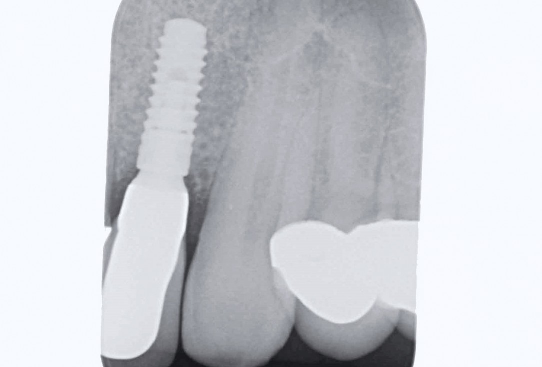
18/19 - X-ray at final restorationBone augementation with maxresorb® - Dr. R. Cutts
-
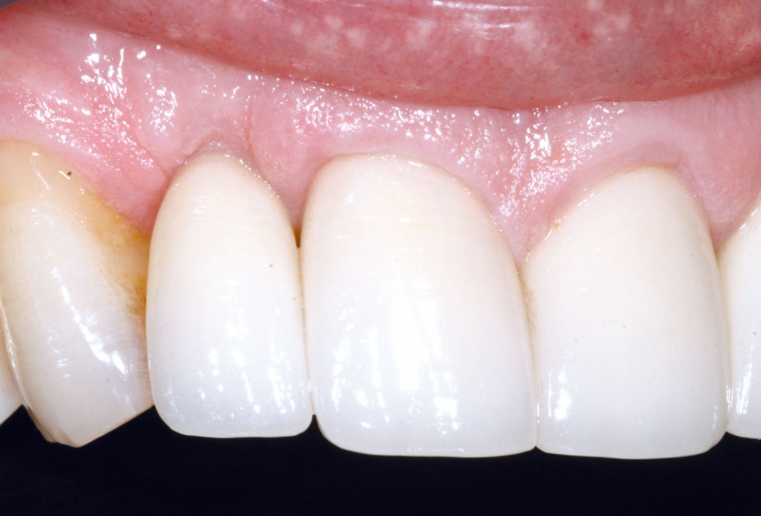
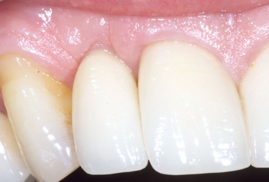
19/19 - Final result 1 year after implant placementBone augementation with maxresorb® - Dr. R. Cutts

Initial Orthopantomograph X-Ray

DVT image showing the reduced amount of bone available in the area of the mental foramen

X-ray shows a 3-dimensional periondontal defect

X-ray control showing initial situation

Surgical presentation of the alveolar ridge with reduced amount of horizontal bone available

Pre-operative x-ray

X-ray control before tooth extraction

DVT image demonstrating horizontal and vertical amount of bone available

Surgical presentation of the alveolar ridge with reduced amount of horizontal bone available

DVT control after sinusitis surgery, residual bone height 1 mm

Clinical situation before extraction

DVT control after sinusitis surgery, residual bone height 1 mm
