Augmentation of the keratinized tissue in the posterior maxilla with mucoderm®- Dr. Dr. A. Stricker
-
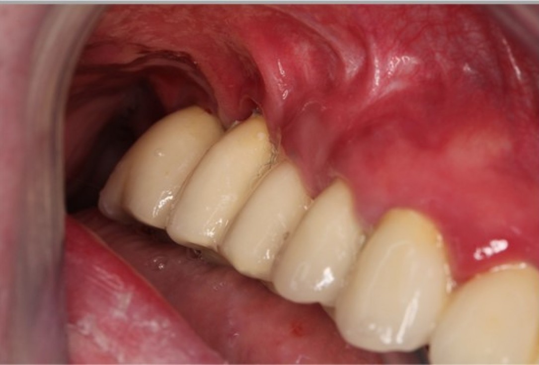
1/7 - Initial clinical situation with lack of keratinized tissueAugmentation of the keratinized tissue in the posterior maxilla with mucoderm®- Dr. Dr. A. Stricker
-
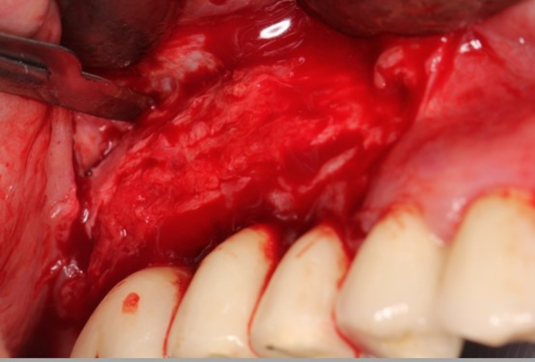
2/7 - Preparation of a mucosal flapAugmentation of the keratinized tissue in the posterior maxilla with mucoderm®- Dr. Dr. A. Stricker
-
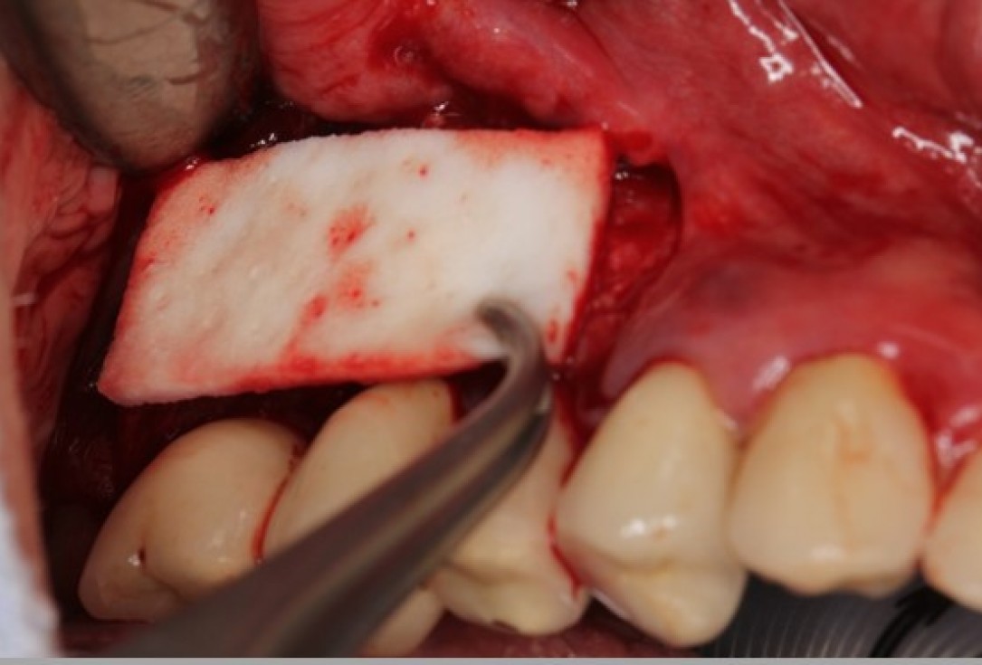
3/7 - Adaptation of mucoderm® to the recipient siteAugmentation of the keratinized tissue in the posterior maxilla with mucoderm®- Dr. Dr. A. Stricker
-
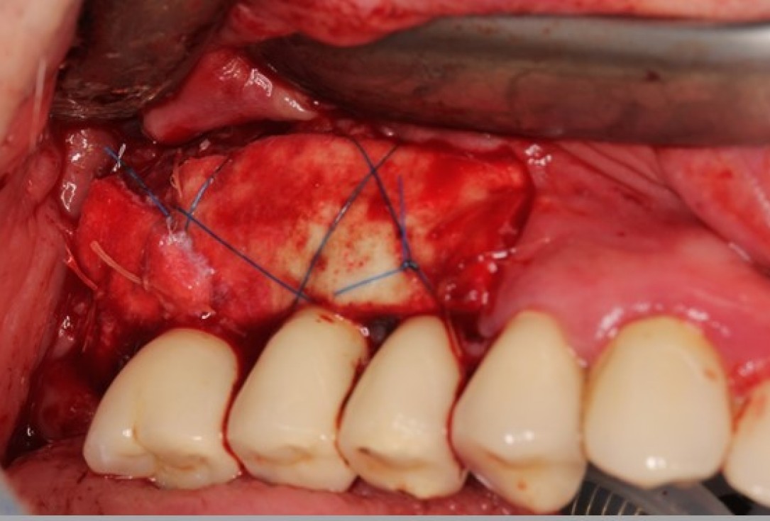
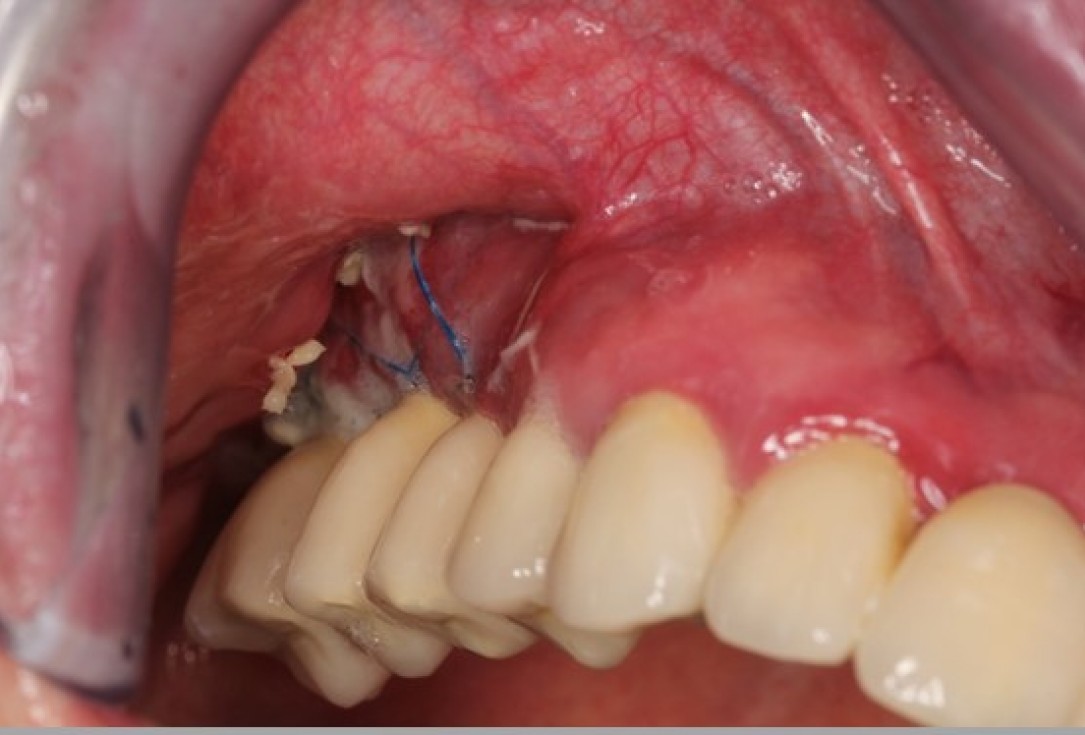
4/7 - mucoderm® sutured tightly to the exposed periosteum and open healingAugmentation of the keratinized tissue in the posterior maxilla with mucoderm®- Dr. Dr. A. Stricker
-
5/7 - Clinical situation at 2 weeksAugmentation of the keratinized tissue in the posterior maxilla with mucoderm®- Dr. Dr. A. Stricker
-
6/7 - Clinical situation at 4 weeksAugmentation of the keratinized tissue in the posterior maxilla with mucoderm®- Dr. Dr. A. Stricker
-
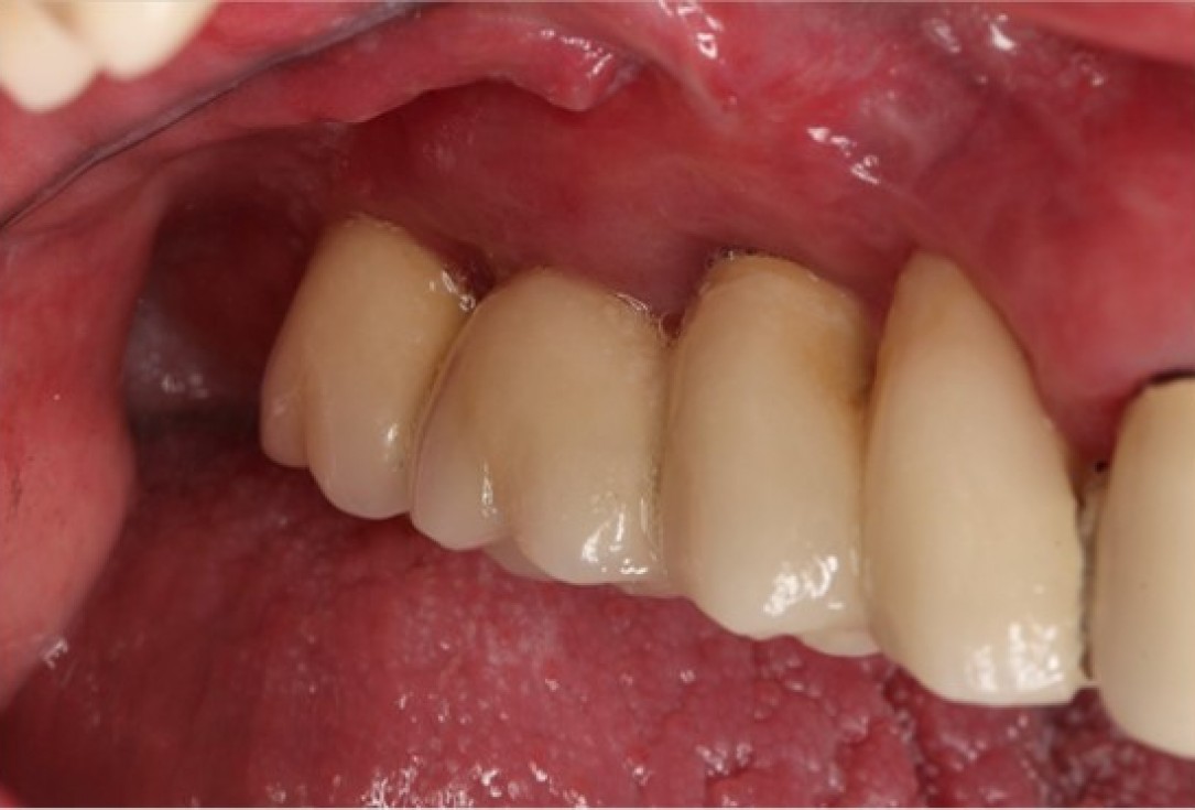
7/7 - Clinical outcome at 3 months with a new band of attached gingivaAugmentation of the keratinized tissue in the posterior maxilla with mucoderm®- Dr. Dr. A. Stricker

Initial clinical situation with loss of attached gingiva around the implants and shallow vestibule

Preoperative situation – Maxillary defect in area 14-16 (loss of implant 16 due to periimplantitis, tooth 14 extracted recently and area 15 already edentulous for a while)

Insufficient keratinized mucosa and extremely shallow vestibule on the maxilla

Pre-operative clinical situation: changed color in the gingiva in the front maxilla

Lack of sufficient keratinized mucosa following extensive horizontal ridge augmentation

Clinical view 8 weeks after extraction of teeth 25 and 26
