Flap surgery in conjunction with a collagen matrix

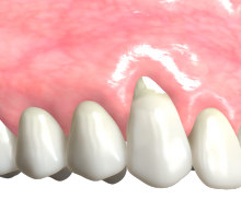
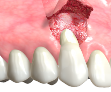
  • Gingival recession at the upper canine. Healthy interdental tissues.
  • Flap preparation. Incisions according to the coronally advanced flap (CAF) technique.
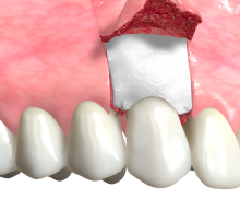
  • Trimmed and rehydrated mucoderm® matrix placed over the exposed root and fixed by sutures.
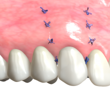
  • Repositioning of the flap in coronal direction and suturing. Complete coverage of the mucoderm® matrix.
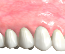
  • Resolution of the defect. Complete root coverage.
Miller class I (recession does not extend to the mucogingival junction (MGJ)) and Miller class II gingival recessions (recession reaches or goes beyond the MGJ) are characterized by healthy interdental tissues and have a good prognosis in terms of complete root coverage. Based on the soft tissue situation, a collagen matrix (mucoderm®) may be used to cover exposed roots as an alternative to the autologous subepithelial connective tissue graft (CTG) [1, 2]. Specifically, for thin biotypes (gingival thickness ≤ 1.0 mm) a volume-supporting approach like the use of mucoderm® is indicated.

Flap techniques

mucoderm® can be used in conjunction with all state-of-the-art mucogingival surgical procedures to treat isolated or adjacent multiple gingival recessions including coronally-advanced flap procedures and tunnel techniques.

Coverage of the collagen matrix

Make always sure to cover the mucoderm® matrix completely by the repositioned flap when used in recession coverage procedures. The collagen matrix requires nutrient and blood supply from the overlying soft tissue when placed on avascular root surfaces. Early exposure may lead to soft tissue graft failure.

Immobilization

To avoid micro-movements and to ensure a close contact to the periosteal wound bed, mucoderm® should be sutured to the intact periosteum when a split flap is prepared. Single button or cross sutures may be used; the use of resorbable sutures is recommended. If the tunnel technique is used, the collagen matrix can be fixed at the level of the cemento-enamel junction of each tooth using sling sutures.

Rehydration

To allow for optimal flexibility and adaptation to the root surfaces, rehydrate the mucoderm® matrix in sterile saline solution or patient blood for 5 to 10 minutes.

Recession coverage with mucoderm® (Prof. Dr. Dr. A. Kasaj)

mucoderm® is a natural type I/III collagen matrix originating from porcine dermis that undergoes a multi-stage purification process, which removes all potential immunogenic substances. The remaining matrix is a membrane with a high interconnected porosity that consists of collagen and elastin [3, 4]. mucoderm® promotes the revascularization and fast soft tissue integration and is a valid alternative to the patient’s own connective tissue [1, 2].

After application, the patient’s blood infiltrates the mucoderm® graft through the three-dimensional soft tissue network, bringing host cells to the soft tissue graft surface and triggering the revascularization process. The collagen network serves as a scaffold for ingrowing blood vessels and cells like fibroblasts, thus supporting a fast revascularization and tissue integration. While collagen is produced by the adhering cells, the matrix is gradually degraded and finally replaced by host tissue [5].

In specific indications the use of an autologous graft may be recommended, e.g. if a very thin flap is prepared or a tension-free flap closure cannot be achieved or a considerable increase in keratinized width is required.


[1] Cosgarea et al. Quintessence Int. 2016;47(9):739-47
[2] Cieślik-Wegemund et al. J Periodontol. 2016 Dec;87(12):1436-1443
[3] Pabst et al. J Periodontal Res. 2014 Jun;49(3):371-81
[4] Pabst et al. Clin Oral Investig. 2015 Mar;19(2):561-4
[5] Rothamel et al. Head Face Med. 2014 Mar 27;10:10