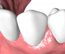
Ridge preservation with dPTFE membrane

  • Ridge preservation with dPTFE membrane
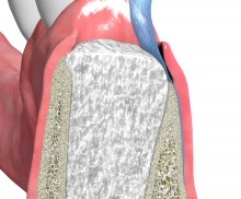
    Deficient bone wall.
  • Ridge preservation with dPTFE membrane
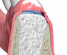
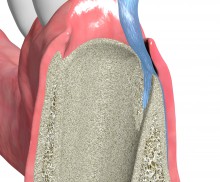
    dPTFE membrane placed to cover the bone defect.
  • Ridge preservation with dPTFE membrane
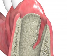
    Alveolar socket grafted with bone substitute material.
  • Ridge preservation with dPTFE membrane
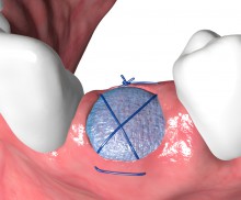
    Bone grafting material covered with dPTFE membrane. The membrane is left exposed for open healing and stabilizied by sutures.
  • Ridge preservation with dPTFE membrane
    dPTFE membrane left exposed to the oral cavity.
  • Ridge preservation with dPTFE membrane
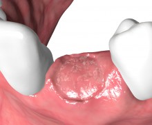
    Situation after membrane removal.
  • Ridge preservation with dPTFE membrane
    Implant-supported restoration.
Alveolar sockets with partially broken or missing bone walls can be regenerated using a high-density PTFE (dPTFE) membrane in an open healing procedure [1, 2, 3]. Due to their small pore size dPTFE membranes act not only as barrier against epithelial downgrowth but also against bacterial penetration, and can therefore be left in place for open healing in socket or ridge preservation. It enables maintenance of the soft tissue architecture and contours since no primary wound closure is required. Due to the missing flap closure, the mucogingival line will not be displaced and the attached/keratinized gingiva will be preserved.

Application & Fixation

To ensure membrane stability and protection of the bone grafting material, permamem® should be placed in such a way that the membrane extends 3-4 mm beyond the edges of the bone defect. On the buccal aspect a mucoperiosteal flap is prepared and permamem® is placed to cover the bone defect completely. Orally, a small mucoperiosteal pocket is prepared extending 3-4 mm beyond the edges of the bone defect, which used to insert and immobilize the membrane. After plane adaptation of the membrane over the socket, permamem® should be immobilized by sutures (horizontal mattress sutures). A minimum distance of 1 mm to the adjacent teeth should be maintained.

Removal

permamem® should be removed after 3-4 weeks. This will provide sufficient time for the formation of the blood clot and a provisional matrix of woven bone in the alveole, which is the basis for the bony regeneration.

permamem® left in place for open healing - Dr. P. Di Capua

Due to its dense structure permamem® acts as an efficient barrier against bacterial and cellular penetration, and may therefore be left in place for open healing in socket or ridge preservation. In ridge preservation the membrane is placed to fully cover the socket and the deficient bone walls, and is stabilized by sutures or pins. After the healing time, permamem® can easily be removed with a pair of tweezers in a non-surgical way. The non-surgical removal of the membrane after the healing time omits the need for big surgical incisions (vertical releasing incisions), thus improving aesthetics and patient comfort. After removal, the primary healing process and the reepithelialization of the regenerating soft tissue will be completed within one month.

In order to promote the osseous regeneration, the socket should be filled with an osteoconductive bone grafting material that acts as a space holder and scaffold for precursor cells; these can then migrate into the socket and defect and mature into osteoblasts, thus triggering the construction of new bone matrix. To achieve this, a mixture of cerabone® and maxgraft® is an excellent option. While the bovine bone ensures a long-term volume stability of the grafted site, the allogenic granules consisting of mineralized collagen promote a fast integration and remodeling, thus supporting the formation of strong, vital bone. As an alternative, cerabone® or the synthetic maxresorb® granules may also be applied individually, particularly if an extended healing time of at least 5–6 months is anticipated.
[1] Carbonell et al. Int J Oral Maxillofac Surg. 2014 Jan;43(1):75-84.
[2] Bartee BK, Compend Contin Educ Dent. 1998 Dec;19(12):1256-8, 1260, 1262-4.
[3] Hoffmann et al. J Periodontol. 2008 Aug;79(8):1355-69.